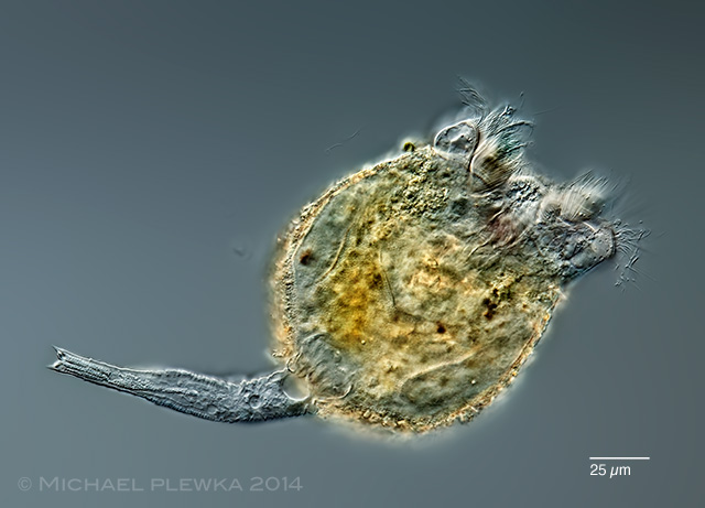
| Brachionus angularis; dorsoventral view of specimen from (2) |
| |
 |
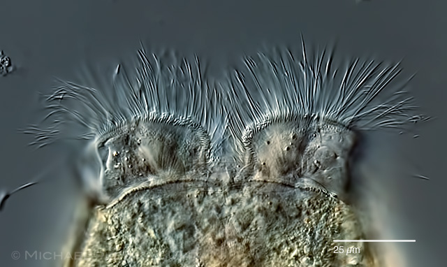 |
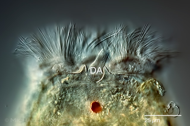
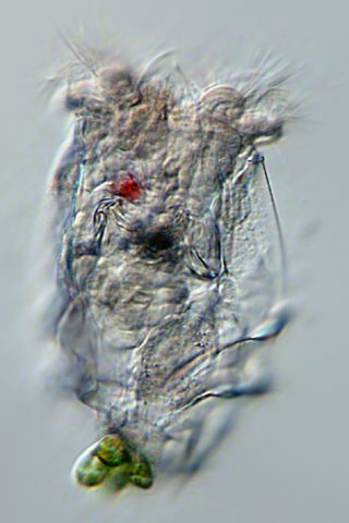
| Brachionus angularis; two aspects of the anterior part of a specimen from (2). Upper image: shows the notch between the two dents in the dorsal part of the anterior lorica where the dorsal antenna protrudes. Lower image: anterior part of the ventral lorica and the interrupted circumapical band of the corona. |
| |
 |
| Brachionus angularis; ventral view. Focus plane on the foot opening of the lorica. (1) |
| |
 |
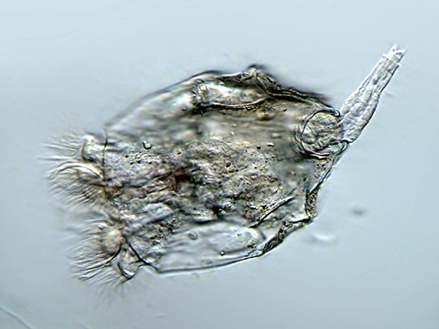
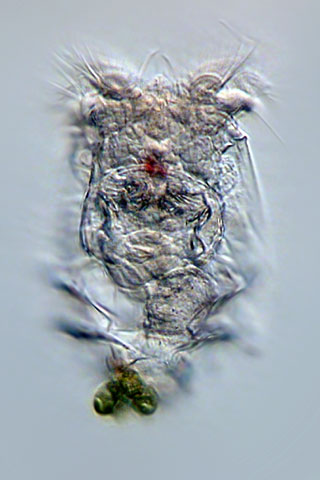
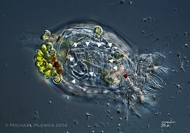
| Brachionus angularis; dorsoventral view, focus plane on the cerebral eyespot, the trophi and the foot. (1) |
| |
 |
| Brachionus angularis; lateral view. The lorica has some transversal notches and a paired lateral notch. (1) |
| |
 |
| Brachionus angularis; dorsoventral view.The right one of the lateral notches is visible. (1) |
| |
  |
| Brachionus angularis; lateral notches in the lorica (1) |
|
| |
 |
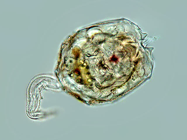
| Brachionus angularis; specimen from (2) with resting egg. |
| |
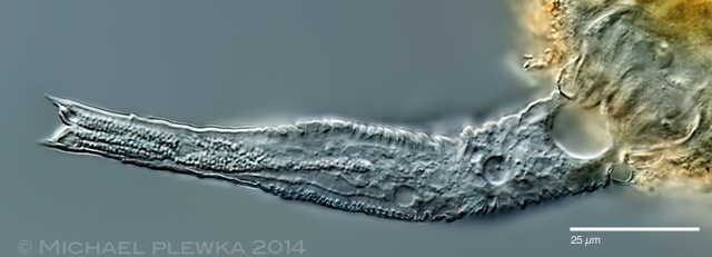 |
| Brachionus angularis; specimen from (3); crop of the above image, detail of the foot. |
| |
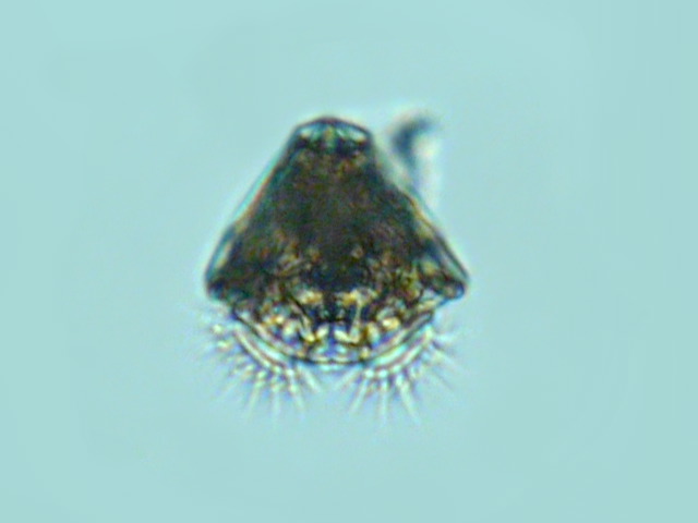 |
| Brachionus angularis; frontal view. (2) |
| |
| |
 |
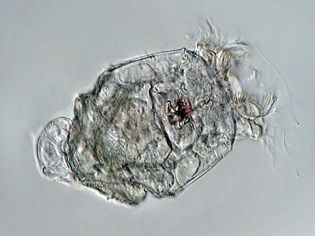
| Brachionus angularis; with several parasites: the euglenid flagellate Colacium sp. as epibiont on the lorica and fungal hyphae are visible in the pseudocoel of this specimen. (2) |
| |
 |
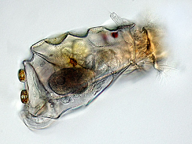
| Brachionus angularis; specimen with amictic egg and the euglenid flagellate Colacium sp. as epibiont on the lorica. Also visible is a peritrich ciliate. (3) |
| |
| |
| |
|
|
| |
| |
| |
| Location: Gevelsberg, Grünes Klassenzimmer (pond) (1); pond IG-Metall-Bildungszentrum, NRW (2;3); |
| Habitat: plankton (1,2,3) |
| Date: 31.05.2009 (1); 07.07.2007 (2); 13.06.2018 (3) |
|
|