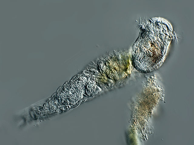
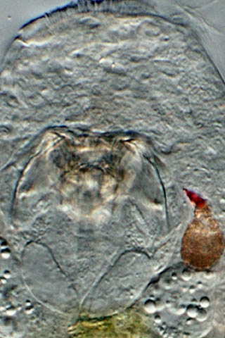
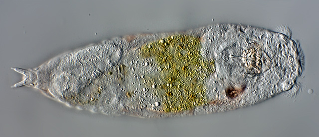
| Notommata groenlandica, ventral view. Focal plane on the conspicuous bulging corona. |
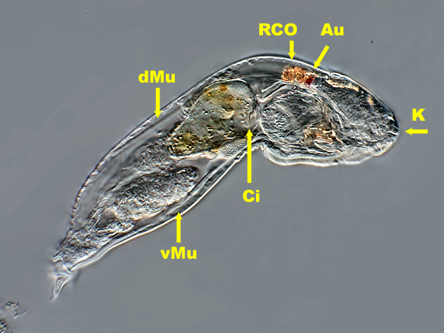 |
| Notommata groenlandica, lateral view. The retrocerebral organ (RCO) has orange inclusions, a dorsal muscle (dMu) leading from the RCO to the back can act as retractor for the head. A ventral muscle (vMu) is also visible. Long cilia (Ci) extend from the oesophagus into the stomach, as is also seen in the video. The head bears a small rostrum, beneath is a strongly refractive body (K, arrow). |
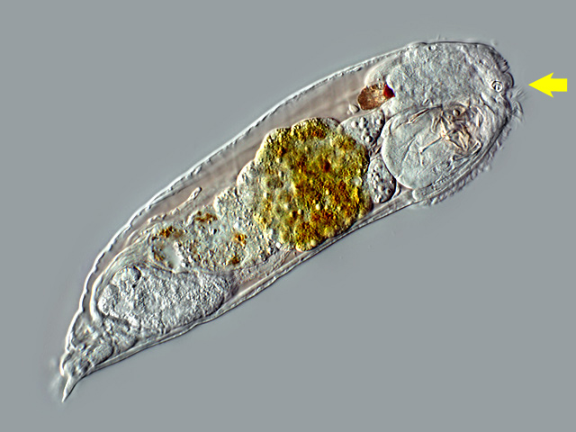 |
| Notommata groenlandica, lateral view of an older specimen, slightly compressed by the cover slide. In contrast to young specimen the humor of older ones is reddish. The arrow points to the refractive body which is kidney-shaped. |
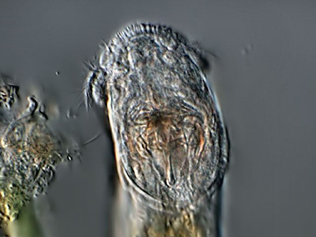 |
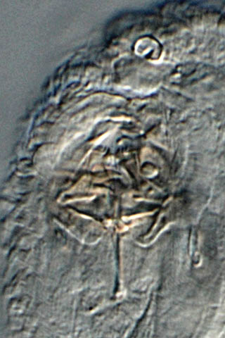
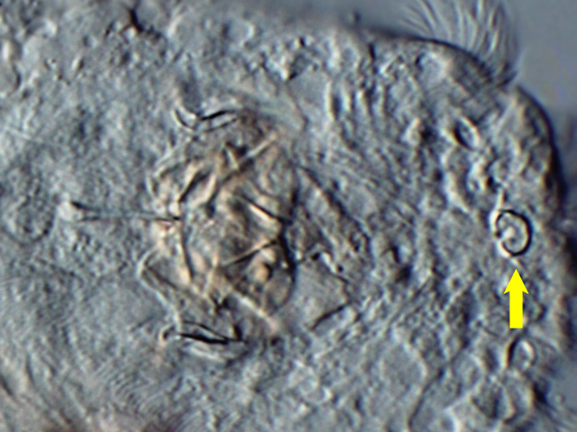
| Notommata groenlandica: head, focal plane on the trophi |
 |
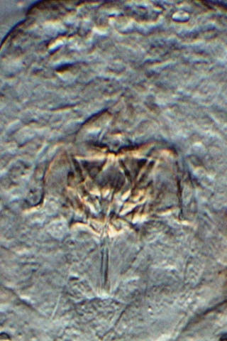
| Notommata groenlandica: ventral view, slightly compressed by coverslide. Focal plane on the trophi and the refracive kidney-shaped body. |
 |
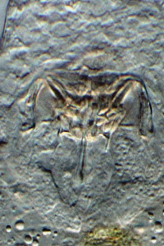
| Notommata groenlandica: crop from the above image. |
|
|
| Notommata groenlandica: 4 aspects of the trophi |
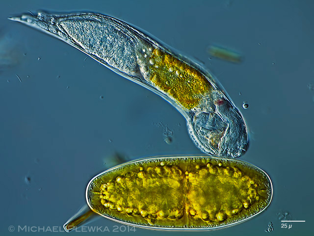 |
| Notommata groenlandica: specimen from (2). Though this image does not document that the trophi can be thrusted out like some Cephalodella-species and Dicranophorids do, it shows the potential function of this behavior: this specimen is trying to open a Netrium-cell, which is a potential prey. |
| Fundort: Wahner Heide bei Köln (1); Gieringer Moor, Tirol, Austria (2) |
| Habitat: Sphagnum-bog (1), (2) |
| Date: 08.05.2010; 27.06.2014 (2) |
|
|