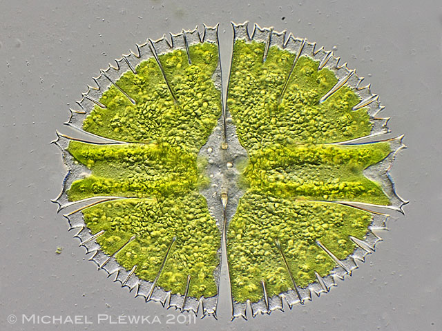
| Micrasterias rotata, specimen from (2). |
| |
 |
| Micrasterias rotata, center of the cell; focal plane on the ridges of the two chloroplasts. The arrowheads point to some of the dictyosomes in the cell plasma. (2) |
| |
 |
| Micrasterias rotata, another specimen, center of the cell; focal plane on the condensed RNA of the nucleolus. The chloroplast seems to be filled with raspberry-like structures (the arrow marks one of them) which are considered to be the stacks of thylakoids (see the image below). (2) |
| |
 |
| Micrasterias rotata, crop of the above image showing granules of thylakoids. (2) |
| |
 |
| Micrasterias rotata, when water under the coverslide evaporates and the cell gets compressed not only lipid droplets start to leak at the tips of the cell but also some structured buckyball-like bodies (arrows, see below) whih may also be dictyosomes. (2) |
| |
  |
| Micrasterias rotata, two images showing the Fulleren-like structures ( ?dictyosomes? )(2) |
| |
| |
 |
| Micrasterias rotata, specimen from (1) |
| |
| |
| |
|
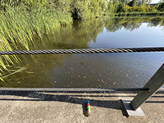
Location (2): Henrichsteich, Hattingen, NRW, Germany, pond |
 |
| |
| Habitat (2): plankton-sample with algae and detritus (click image to enlarge >>>) |
| |
| Date (2): ; 14.03.2023 (2) |
| |
|
|
|
|
|
| |
|
| Location (1): Wahner Heide bei Köln (1); |
| Habitat (1): Spagnum- Probe |
| Date (1): 19.06.2010 |
|