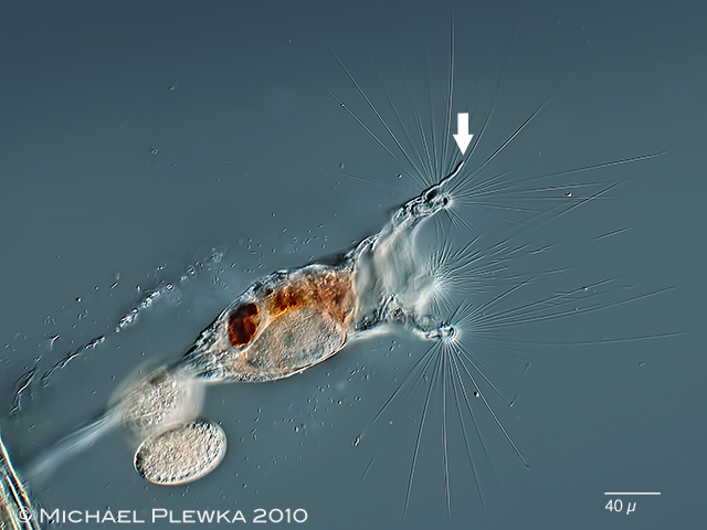
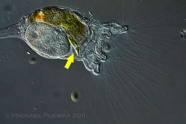
| Collotheca ornata var. cornuta, lateral view, is characterized by a worm-like process at the middle dorsal lobe of the corona (arrow). This is in contrast to Collotheca ornata var. ornata (3) |
| |
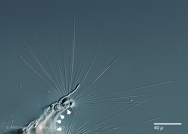 |
| Collotheca ornata var. cornuta, crop of the above image. The arrowheads in this image point to one of the five the interspaces between the tips of the lobes. All five interspaces are free of short cilia. This is in contrast to Collotheca coronetta. It is also fascinating to see that when the corona is extended, these long cilia of the corona become straight like a ruler after they had been totally twisted during contraction. (3) |
| |
 |
| Collotheca ornata, ventral view (3) |
| |
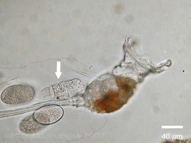 |
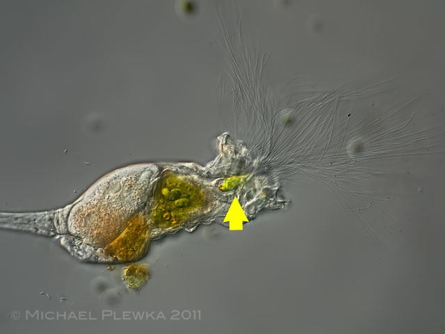
| Collotheca ornata, lateral view, adult female. The mucilaginous sheath, which surrounds the foot and the body base, contains some amictic eggs and a newly hatched juvenile specimen (arrow) which still has red eyespots. The sheath (into which the animal can retract) is secreted by the integument. |
| |
 |
| Collotheca ornata, dorsoventral view of a juvenile specimen. The juveniles swim around and have two red eyespots which disappear when the juveniles settle and the corona begins to grow. |
| |
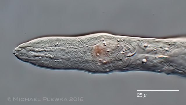 |
| Collotheca ornata, posterior part of the body. Detail of the foot with developing foot glands. |
| |
 |
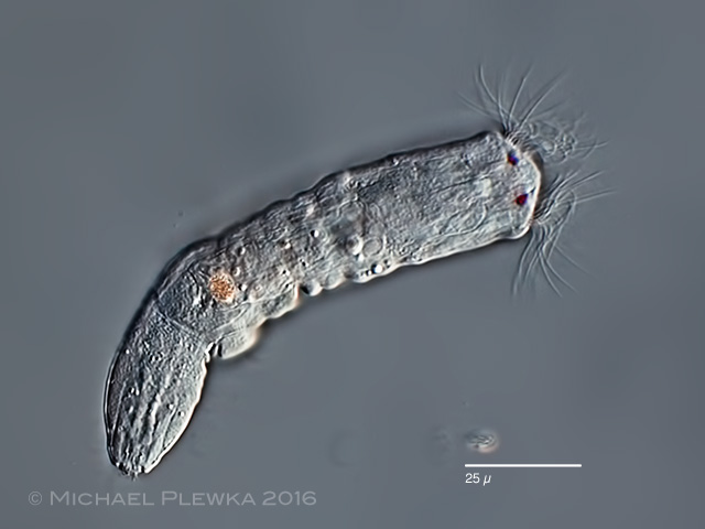
| Collotheca ornata, lateral view, juvenile specimen; the corona is half developed, still with eyespots.The worm-like process is already developed. (3) |
|
| |
 |
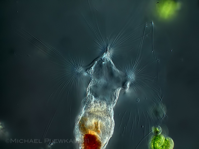
Collotheca ornata, specimen from (2). Since its discovery this critter fascinates by its corona which is partly built from clusters of long cilia. The question arises: what is the function of these cilia? It seems obvious that the cilia might catch the prey, but there are some doubts.
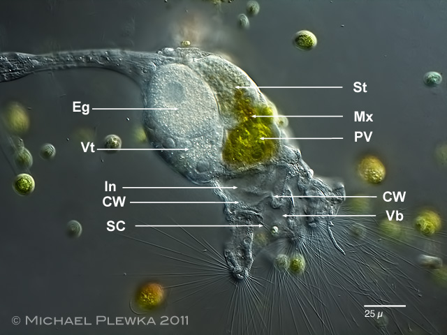
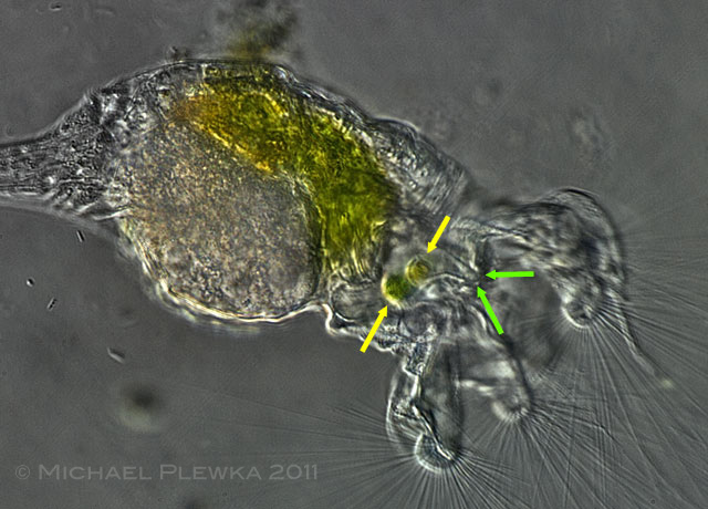
CW: ciliary wreath; Eg: egg; In: infundibulum; Mx: mastax with trophi; PV: proventriculus; SC: sensory cilia; Vt: vitellarium.
|
| The funnel of the "mouth" is divided into two parts: the vestibulum (Vb) and the infundibulum (In) which are separated by a cilia-carrying membrane (ciliary wreath, CW). (3) |
|
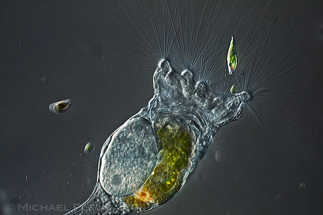 |
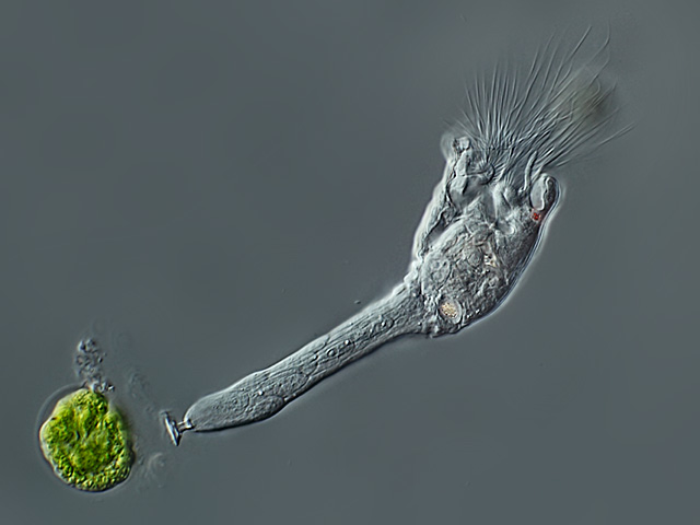
| First I could observe the animal with the corona stretched out. It is always fascinating to see how straight the cilia are. A potential prey, an euglenid algae is visible. (3) |
| |
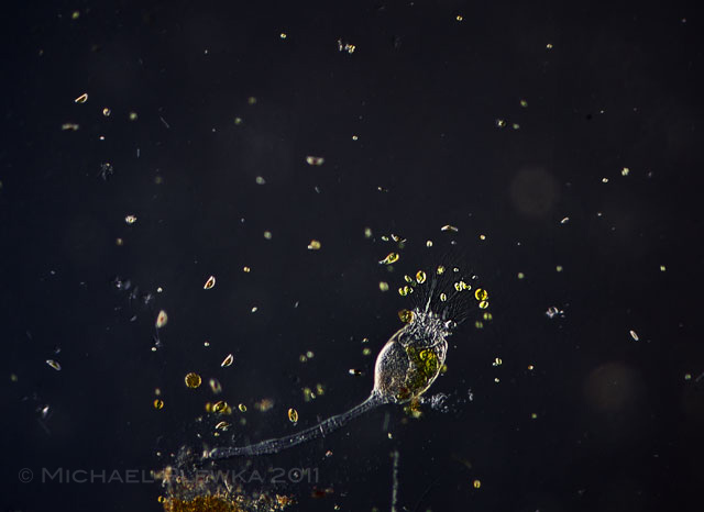 |
| This image shows the distribution of (euglenoid) flagellates, a potential prey around Collotheca. It is obvious that these mobile flagellates are more concentrated around Collotheca. (3) |
| |
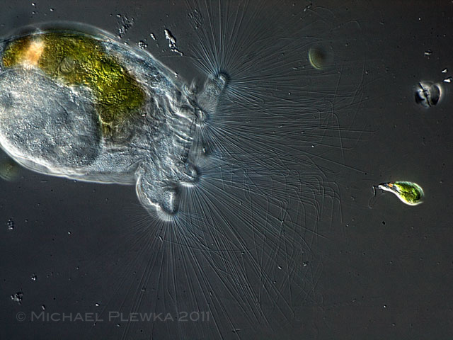 |
| If such a potential prey gets /swims actively into the thicket of these cilia, the cilia start to tremble, and a wavelike movement is creeping along the single cilia; partly the cilia are bent perpendicular. However there is no ingestion of the prey. The algae can swim out of the trap with no effort. The reason maybe that the stomach is so full of diet that the mastax (the trophi are permanently working) is not able to transport additional algae into the stomach. (3) |
| |
 |
| The situation changes when part of the content of the stomach/ intestinum is excreted so that the stomach can take up new diet. If a potential prey (in this case two small euglenoid flagellates) swims actively into the trochus (and probably gets in contact with the sensory cilia), the animal contracts the trochus, the flagellates are pressed into the vestibulum and the mebrane between trochus and vestibulum closes. The prey is pressed into the oesophagus and then transferred into the stomach by the trophi (see also the next two images below). (3) |
| |
 |
 |
| Conclusion: While some authors claim to have observed that the cilia can act as a mechanical trap for potential prey in some Collotheca species, the possibility that -at least- C. ornata uses the cilia (only?) to distribute some chemical (organic) attractant into the water thus attracting mobile algae cannot be excluded. This hypothesis is supported by observations on the behavior of the nearly immobile Collotheca edenta which feeds by just opening the mouth which is fringed with very short cilia, "waiting" for the (mobile) algae like diatoms to actively move into the infundibulum. (3) |
| |
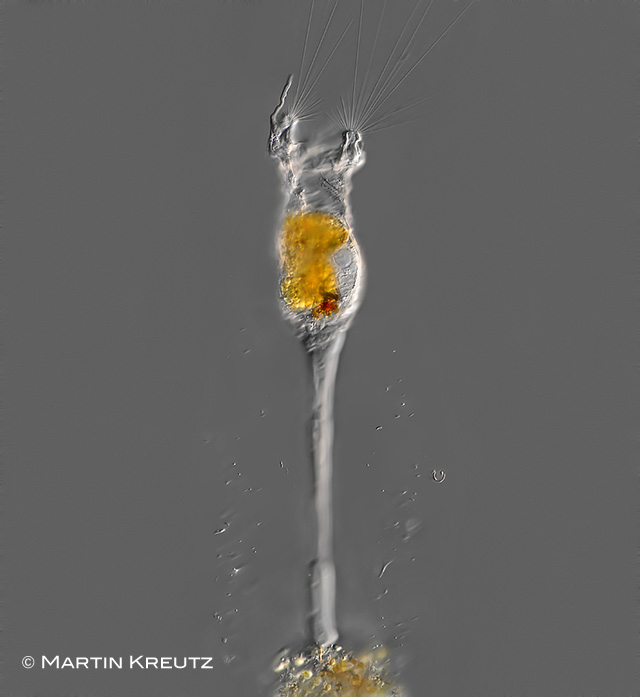 |
| Collotheca ornata cornuta, specimen from (4 ). Image courtesy of Dr. Martin Kreutz, realmicrolife. |
| |
| |
|
|
| |
| |
| Location: Gevelsberg, Grünes Klassenzimmmer, Teich (1); Wahner Heide near Cologne (2); Hattingen, Wodantal, Teich (3); Simmelried near Konstanz (4) |
| habitat: water plants (Potamogeton sp.) (1),(2); water plants (3; 4) |
| Date: 17.04.2007(1); 20.03.2011 (2); 12.06.2010 (3); 13.12.2008 (4) |
|
|