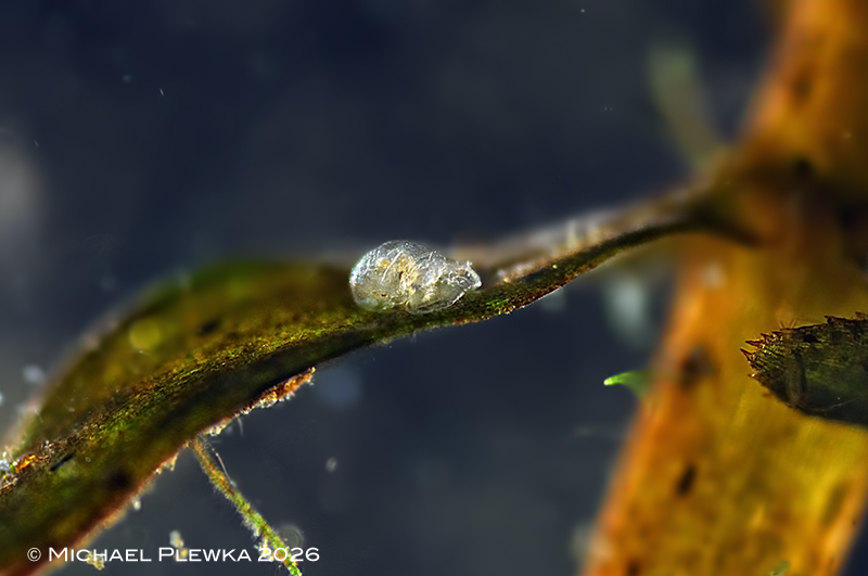
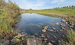
| Cupelopagis vorax: frontal view, on a leaf of the waterweed Elodea sp. (2) |
| |
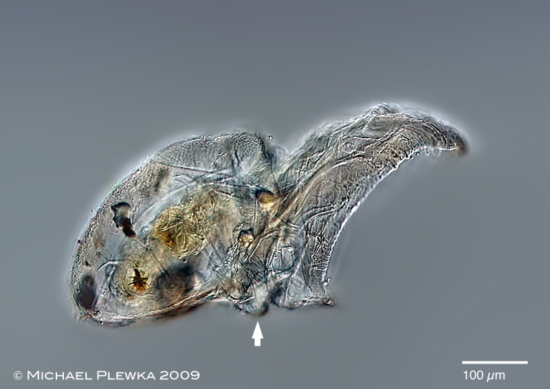 |
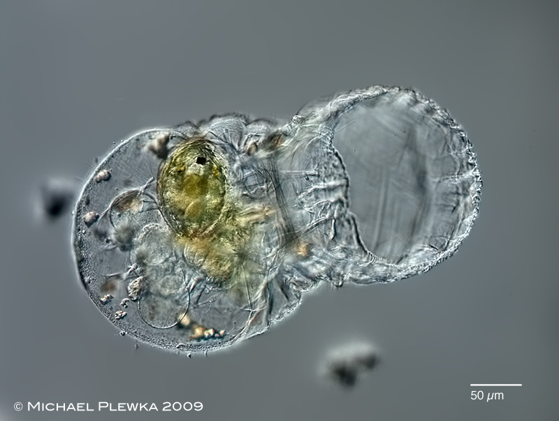
| Cupelopagis vorax: specimen removed from substratun; lateral view, focal plane on the big funnel (infundibulum). The arrow points to the area of the adhesive disc. (1) |
| |
 |
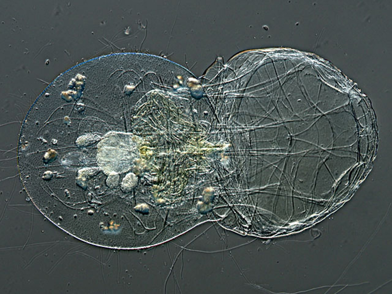
| Cupelopagis vorax: another specimen, ventral view. A green ostracod is visible in the intestinum. Non-digestible remnants of the diet are stored in grains. (1) |
| |
 |
| Cupelopagis vorax: sightly dorsoventrally compessed specimen (by coverslide) showing the muscles. (1) |
| |
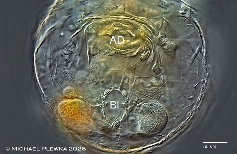 |
| Cupelopagis vorax: trunk, ventral view, focal plane on the (retracted ) adhesive disc (AD) and the bladder (Bl). (1) |
| |
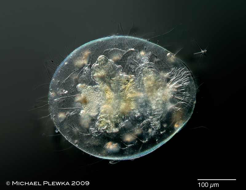 |
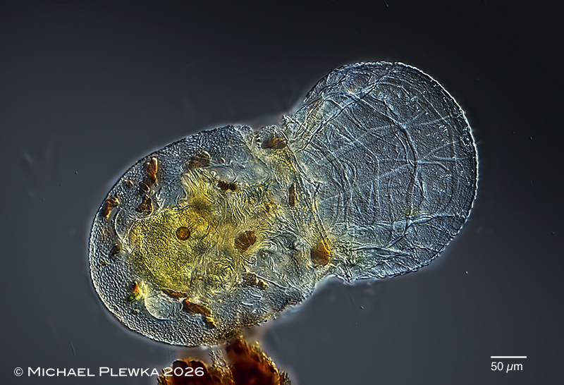
| Cupelopagis vorax: contracted specimen. (1) |
| |
 |
| Cupelopagis vorax: specimen from (2), dorsoventral view. In contrat to all other species of the taxon Euroatoria the corona of this species has no cilia except two ciliated cushions (see image below) |
| |
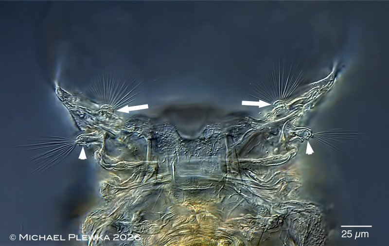 |
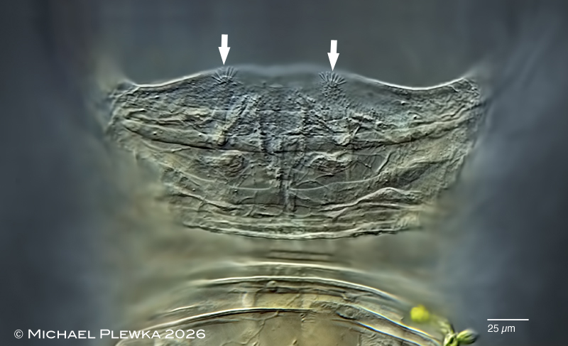
| Cupelopagis vorax: ventral view; focal plane on the lateral antennae (arrowheads) outside and the two ciliated cushions of the pharyngeal sensory organ (arrows) inside the funnel. (2) |
| |
 |
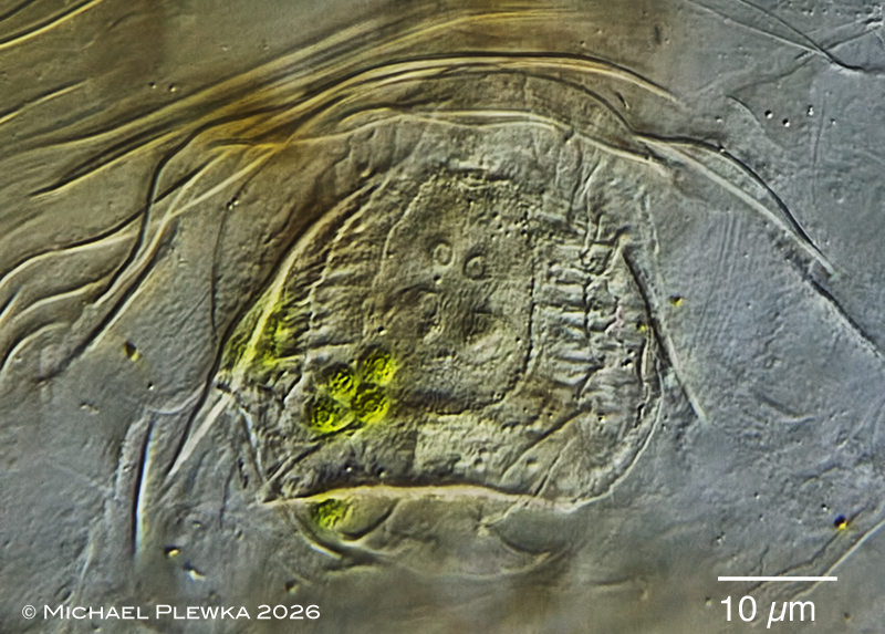
| Cupelopagis vorax: another ciliated structures (arrows) on the ventral side of the rim of the funnel (2) |
| |
 |
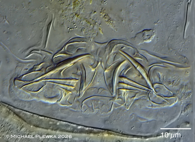 |
| Cupelopagis vorax: digestive system of living specimen; upper image: entrance of the mastax; lower image: uncinate trophi . Both images are stills from a YT-video of the trophi in action >>> (2) |
| |
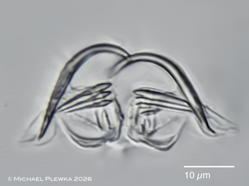 |
| Cupelopagis vorax: uncinate trophi of macerated specimen (2) |
| |
| |
|
|
| |
| |
| location (1): aquarium. the specimens were provided by A. Werth |
| Habitat (1): on the leafs of Nymphaea alba |
| Date: 06.09.2009 (1) |
| |
| |
| |