| Itura viridis, creeping specimen; ventral view. Focus plane on the ventral ciliary field.(2) |
| |
 |
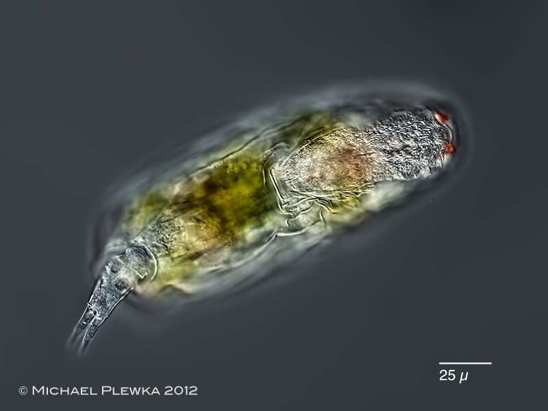
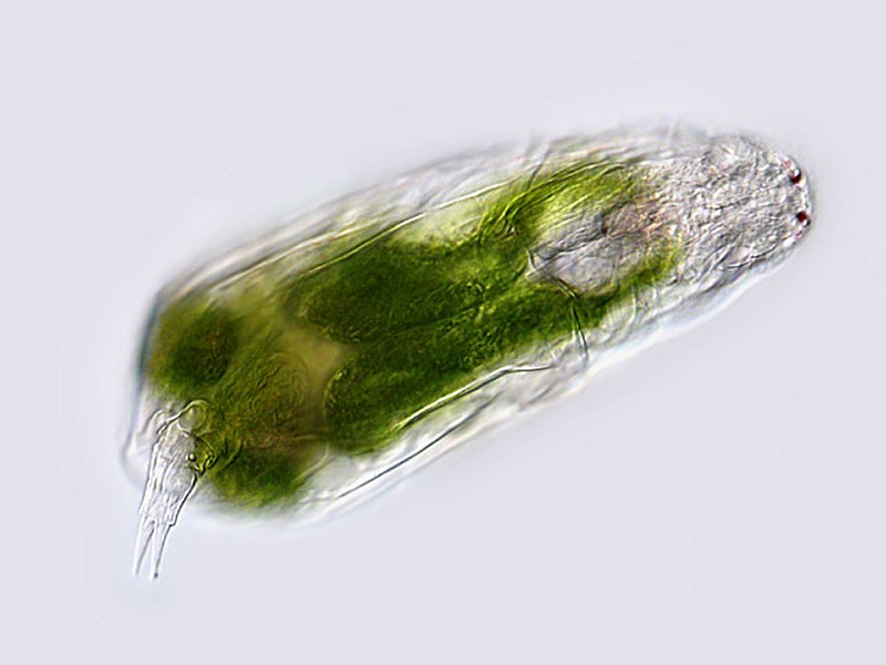
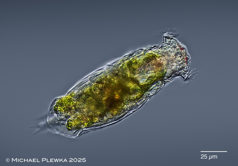
| Itura viridis, a species with lenses in the frontal eyespots (see below). Specimen from (2) |
| |
 |
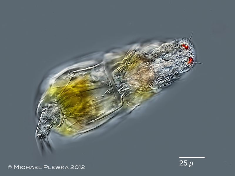
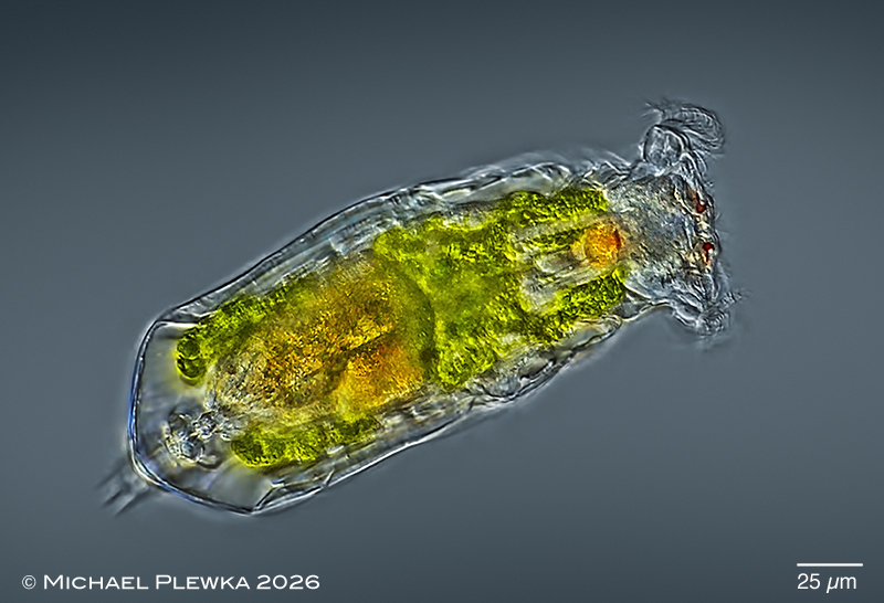
Itura viridis, specimen from (1).
Itura is one of the few rotifer genera that can host algal cells (zoochlorellae). Other rotifer genera with zoochlorellae are: Ascomorpha, Dicranophoroides and Parencentrum. |
| |
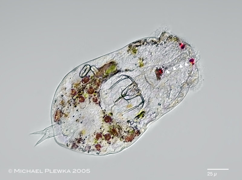 |
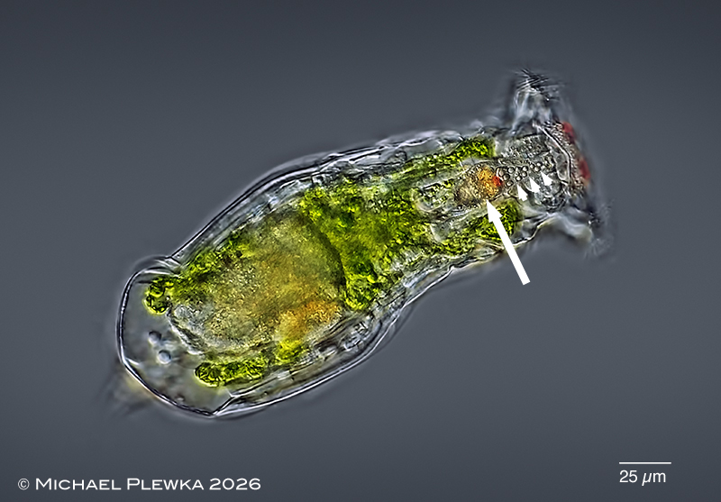
| Itura viridis, heavily compressed specimen from (1) showing several digested algae (brown) in the stomach. 3 paramylon grains of digested euglenid flagellates are visible. Manubria of the trophi with lateral extensions on the outer side (see also images below). The triangles mark the duct of the retrocerebral sac (with red pigment spot) which is relative short in comparison to the duct of Itura aurita. Because of this the retrocerebral sac is located anterior of the trophi. |
| |
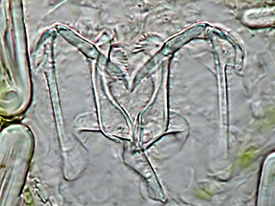 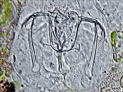 |
| Itura viridis: left: virgate trophi of specimen from (2); right: trophi of specimen from (1). In contrast to Itura aurita the alulae of the rami are lacking the external lamellae. |
|
| |
 |
| Itura viridis, swimming specimen with extended auricles; dorsoventral view. Specimen from (4) |
| |
| |
 |
| Itura viridis, swimming specimen with extended auricles; dorsoventral view. Specimen from (5) |
| |
 |
| Itura viridis, same specimen; focal plane on the retrocerebral sac with red (?eye?) spot (arrow) and duct (arrowheads). (5) |
| |
| |
| |
| |
|
|
| |
| |
|
|
| |
| |
| Location: Wahner Heide ; Köln (3); Hattingen, Wodantal, pond (2) |
| Habitat: puddle (2); detritus (3) |
| Date: 8.6.2009 (2); 04.03.2019 (3) |
|
|