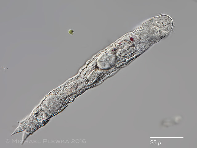
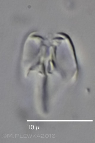
| Pourriotia werneckii dorsoventral view. Visible is the single eyespot which is displaced to one (? right ?) side . Some more red pigment granules are scattered posterior of the eyespot. (1) |
| |
 |
| Pourriotia werneckii, lateral view. Focus plane on the eyespot. (1) |
| |
 |
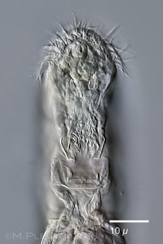
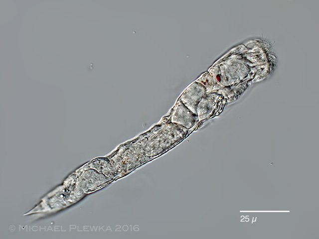
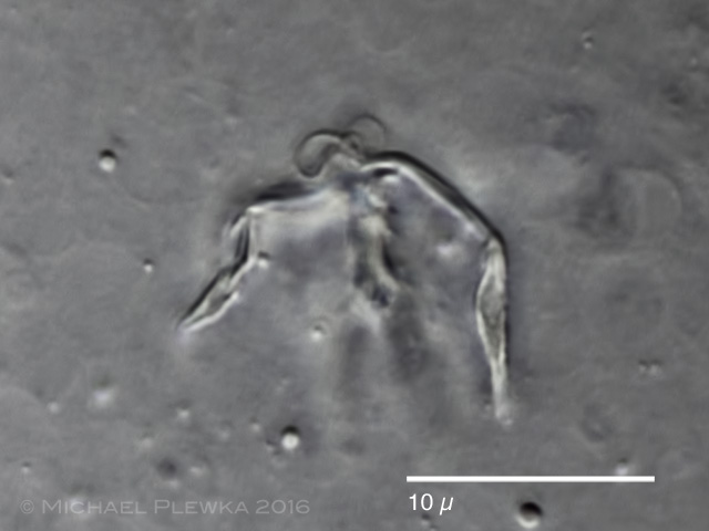
| Pourriotia werneckii; ventral view. (1) |
| |
|
|
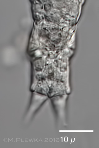
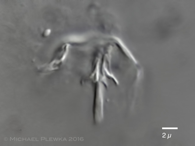
| Pourriotia werneckii; left image: crop of the above image showing the buccal field. Right: foot and toes. (1) |
| |
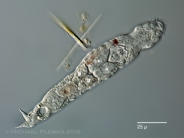 |
| Pourriotia werneckii; showing two pairs of glands labeled " * " , (salivary glands) , and " # " (gastric glands), and the germovitellarium (labelled " ◊ "). (1) |
| |
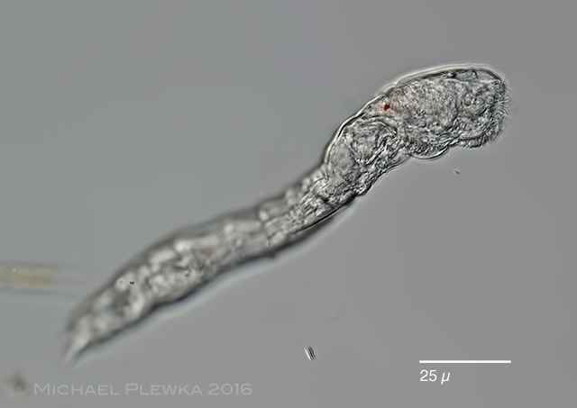 |
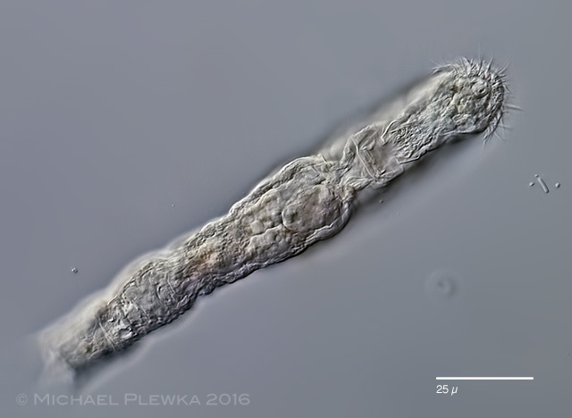
| Pourriotia werneckii; another specimen from the same location; lateral view. (1) |
| |
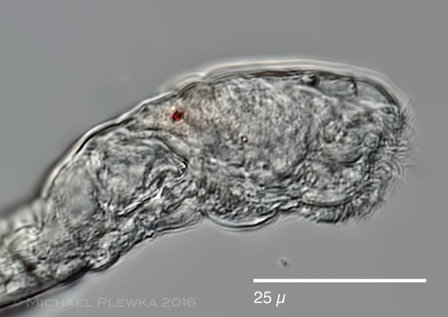 |
| Pourriotia werneckii; crop of the above image. (1) |
| |
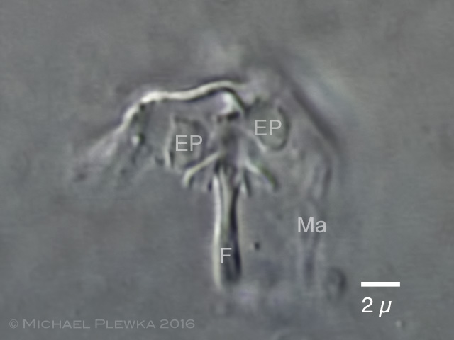 |
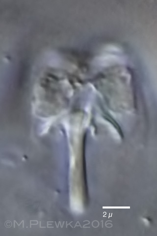
| Pourriotia werneckii; trophi aspect 1 showing the epipharyngeal plates (EP) which have been overlooked by Rousselet. (1) |
| |
 |
| Pourriotia werneckii; trophi aspect 2. |
| |
 |
| Pourriotia werneckii; trophi aspect 3. |
| |
|
|
| Pourriotia werneckii; trophi of another specimen (30.04.). (1) |
|
| |
|
|
| |
| |
| |
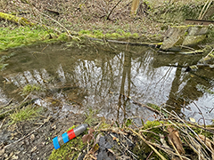
| Location: Hattingen Felderbachtal, Mühlteich. |
| Habitat: Periphyton |
| Date: 24.04.2016; 30.04.2016 |
|
|
|