|
|
| Resticula gelida Harring & Myers 1922 |
 |
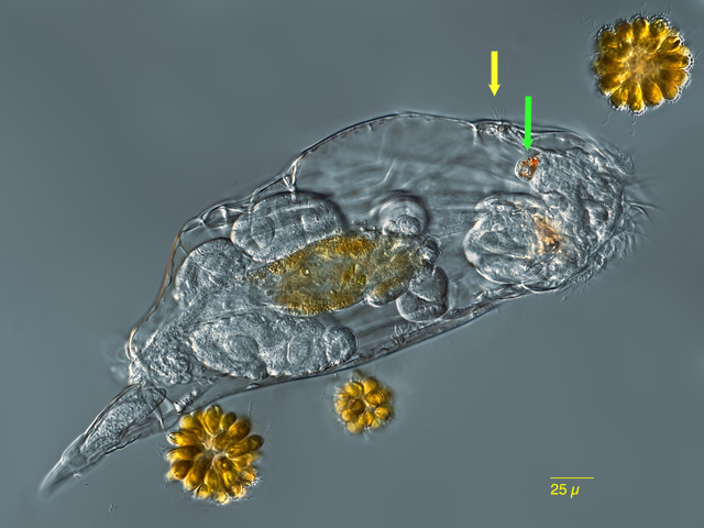
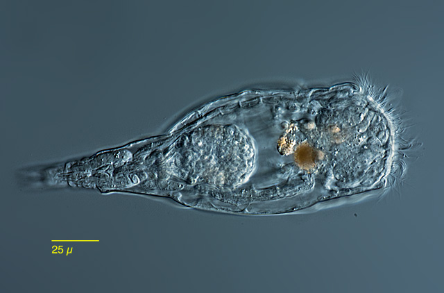
| Resticula gelida, female specimen swimming, dorsoventral view. |
| |
 |
|
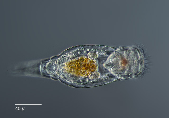
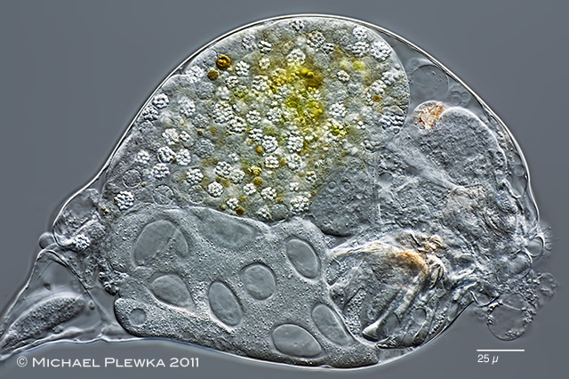
| Resticula gelida, female specimen, lateral view. The dorsal antenna is visible (yellow arrow). The green arrow points to the retrocerebral sac which is filled with orange grains. |
| |
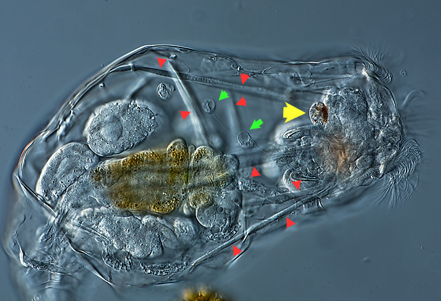 |
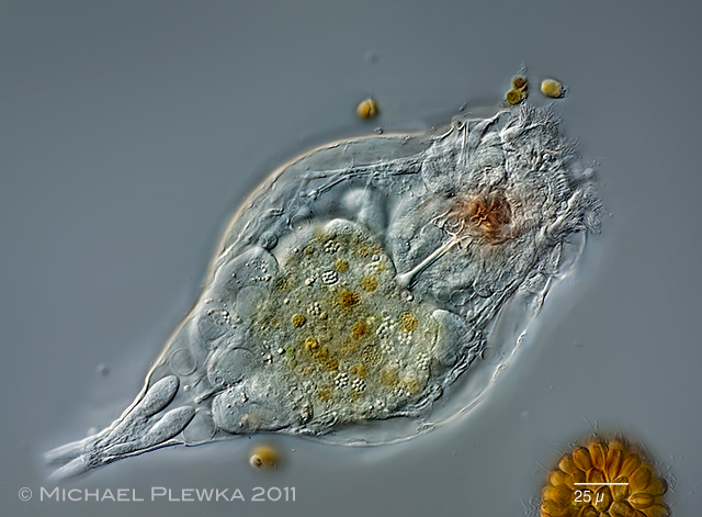
| Resticula gelida, female specimen slightly compressed by the coverslide, dorsoventral view. Yellow arrowhead: retrocerebral sac; red arrowheads point to the longitudinal and transversal muscles; green arrowheads point to amoeboid cells (amoebocytes). These amoebocytes can also be observed in Asplanchna. |
| |
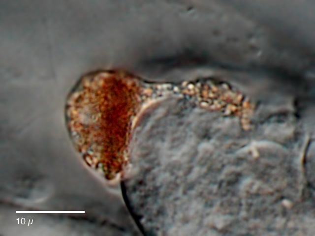 |
| Resticula gelida, female specimen, retrocerebral sac. |
| |
 |
| Resticula gelida, male specimen, dorsoventral view. In contrast to other monogont male rotifers the males of R. gelida are only slightly smaller than the females, but can be distinguished from the latter by the lacking trophi. |
| |
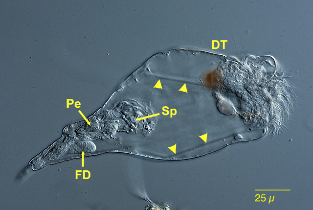 |
| Resticula gelida, male specimen, lateral view; DT: dorsal antenna; Pe: penis; Sp: sperms; FD: foot glands; the arrowheads point to the body musculature. |
| |
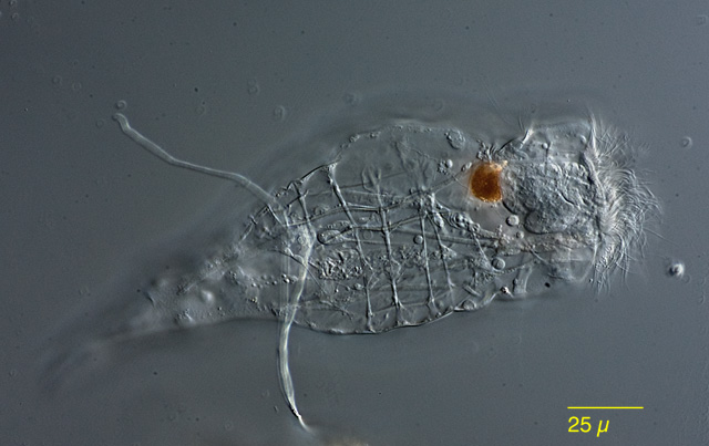 |
| Resticula gelida, male specimen, focal plane on the body musculature. |
| |
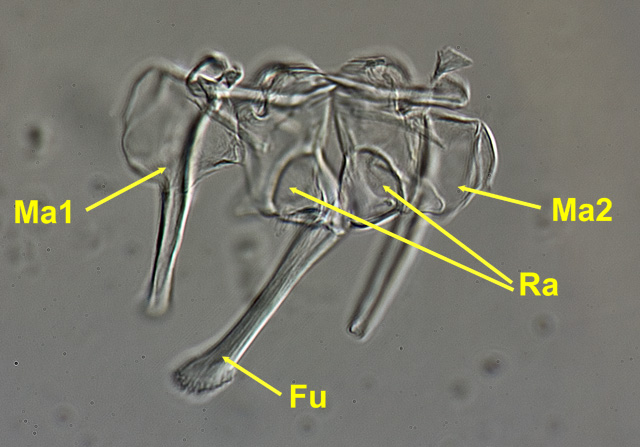 |
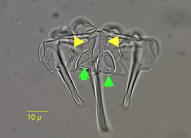
| Resticula gelida, trophi, focal plane on the fulcrum (Fu), manubria (Ma) and rami (Ra). |
| |
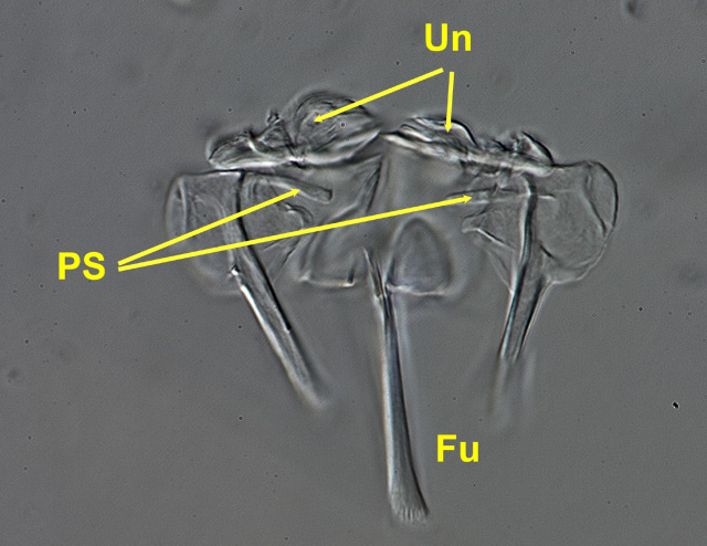 |
| Resticula gelida, trophi, focal plane on the fulcrum (Fu), dentated unci (Un), and pleural rods (PS). |
| |
 |
| Resticula gelida, trophi; the rami have dentations on the inner margins (yellow arrowheads) and are windowed (green arrowheads). |
|
| |
 |
| Resticula gelida, specimen infested by parasites. (1) |
| |
 |
| Resticula gelida, same specimen, compressed by coverslide; lateral view, focus plane on the parasites.(1) |
| |
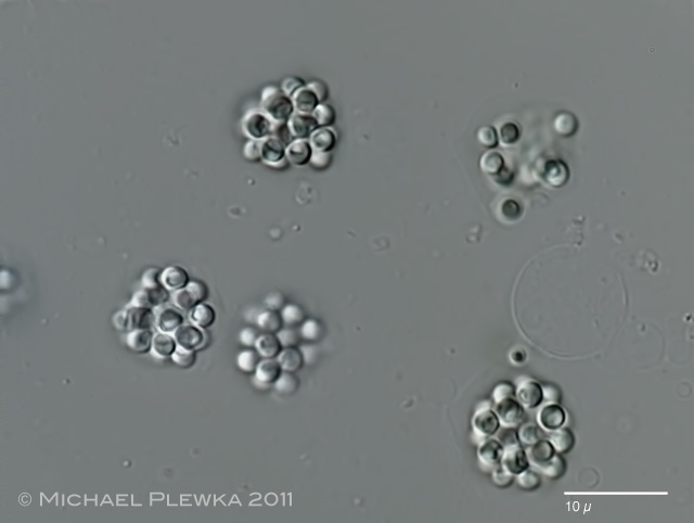 |
| Resticula gelida, parasites.(1) |
| |
| Location: Sprockhövel Hasslinghausen, Hasslinghauser Mulde, pond covered with ice. (1); |
| Habitat: Detritus (1) |
| Date: 25.02.2011 (1); |
|
|
|
|
|
|
|
|