|
|
| Epiphanes brachionus Ehrenberg 1837 |
 |
| Epiphanes brachionus; swimming specimen, dorsoventral view, optical median section. (2) |
| |
 |
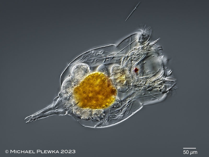
| Epiphanes brachionus, swimming specimen, dorsoventral view. Focal plane on the buccal field. In contrast to Epiphanes senta the shape of the body is more or less angular; the foot is distinct. (2) |
| |
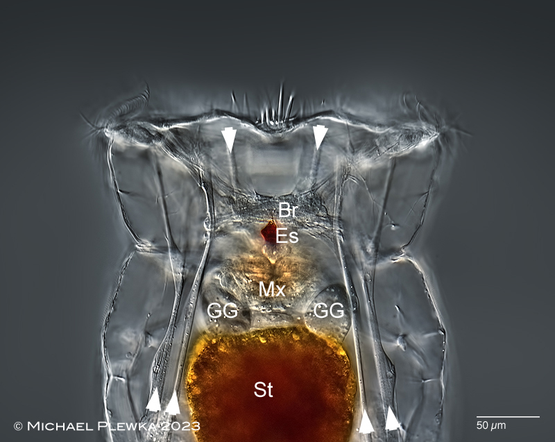 |
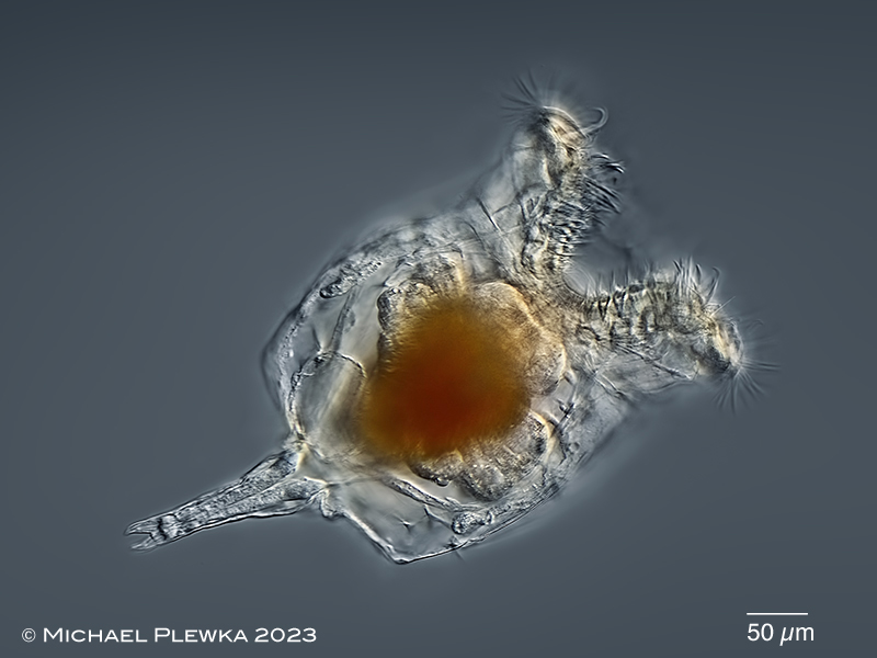
| Epiphanes brachionus, swimming specimen, dorsoventral view. Focal plane on the brain (Br) and the red eyespot (Es), which is in contrast to the colorles eyespot of Epiphanes senta . Also visible are some retractor muscles (arrowheads) and the gastric glands (GG). Mx: mastax; St: stomach. (2) |
| |
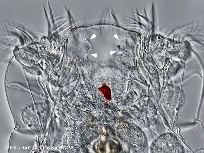 |
| Epiphanes brachionus, head; focal plane on the buccal field. Also visible are the ducts of the RCO (arrowheads). (2) |
| |
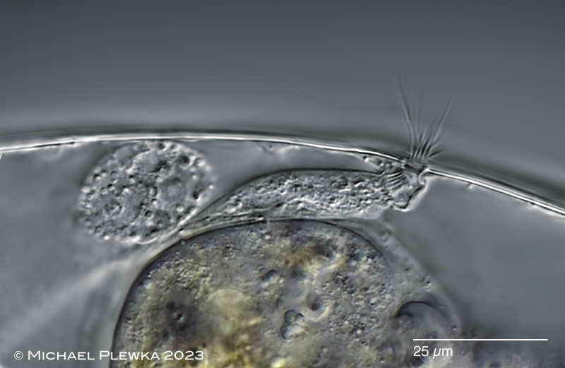 |
| Epiphanes brachionus, right lateral antenna. (2) |
| |
| |
| |
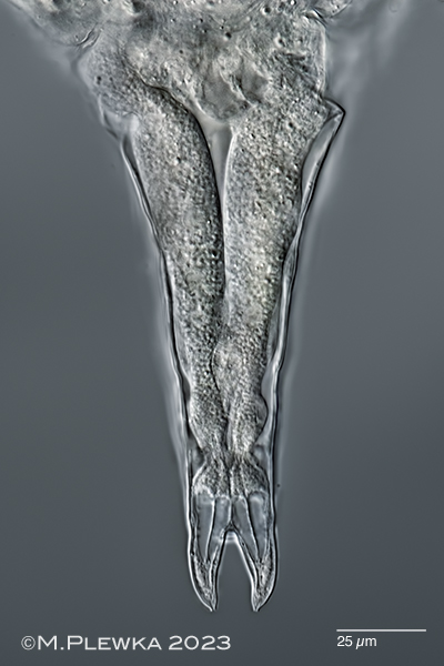 |
| Epiphanes brachionus, foot with footglands. (2) |
| |
| |
  |
|
| Epiphanes brachionus, malleate trophi; left image: focal plane on the unci with four majoer teeth (1). Right: ??epipahryngeal?? structures anterior of the mastax (2). |
| |
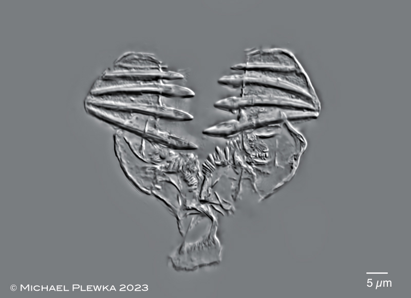 |
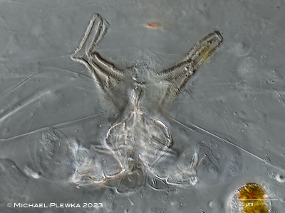
| Epiphanes brachionus, malleate trophi of specimen from (2). |
| |
| |
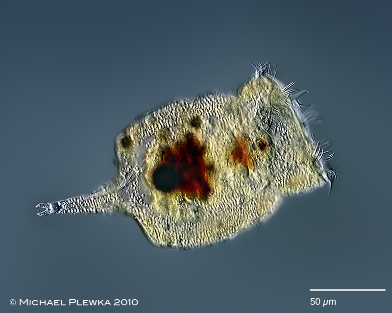 |
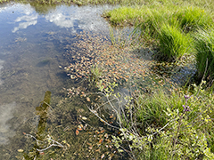
| Epiphanes brachionus , ecology: specimen with endoparasites (1) |
| |
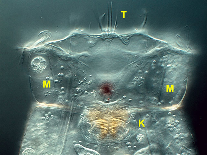 |
| Epiphanes brachionus: head; focal plane on the sensory bristles (T), muscles (M) and trophi (K). (1) |
| |
| |
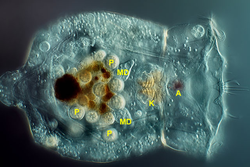 |
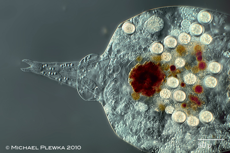
| Epiphanes brachionus , a specimen infected by parasites (P). A: eyespot; MD: gastric glands; K: trophi. (1) |
| |
 |
| Epiphanes brachionus: posterior part of the body and foot infected by parasites. |
| |
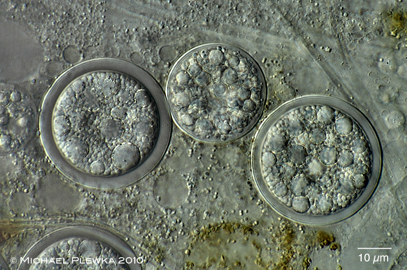 |
| Epiphanes brachionus: close-up of parasites. |
| |
| |
| |
| |
| |
|
|
| |
|
|
|
|
|