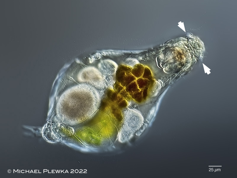
| Notommata copeus: specimen from (4). The specimens of some populations are reported to have large T-shaped retractable auricles (arrowheads); the specimens from this population (3) / (4) (sampled at different times) only showed small auricles. |
| |
 |
| Notommata copeus: the body is often covered with a mucilaginous sheath which is occupied by bacteria. Such mucus layer can also be found in other rotifers such as Proalinopsis caudatus. |
| |
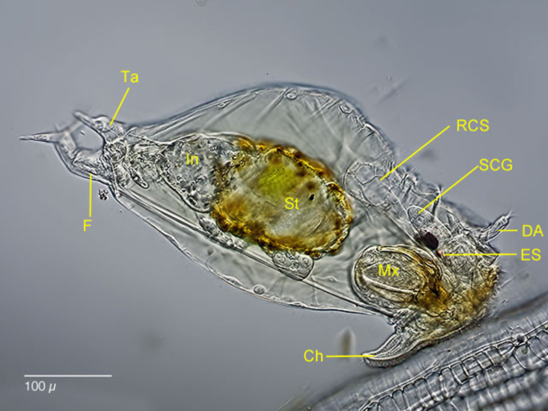 |
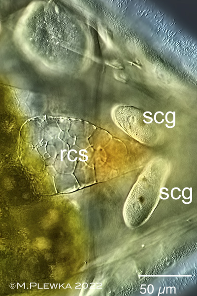
| Notommata copeus: lateral view of a specimen at a leaf of sphagnum. The extreme long an vacuolized retrocrebral sac (RCS) and the long subcerebral glands (SCG) with darg inclusions. Mx: virgate mastax; Ch: "chin" (ciliary field of the wheel organ); DA: dorsal antenna; ES: eyespot; F: foot; In: intestinum; Ta: tail. |
| |
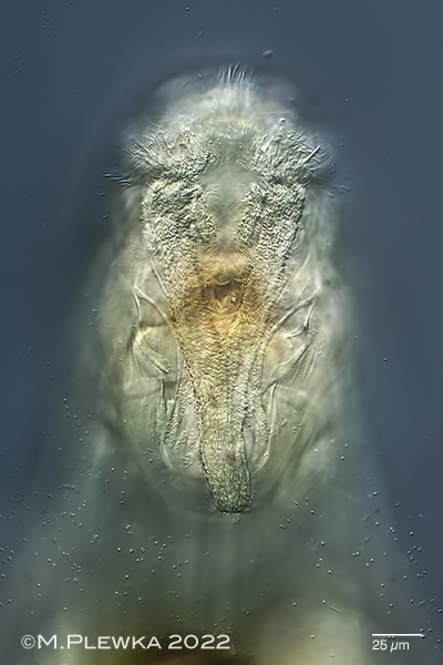 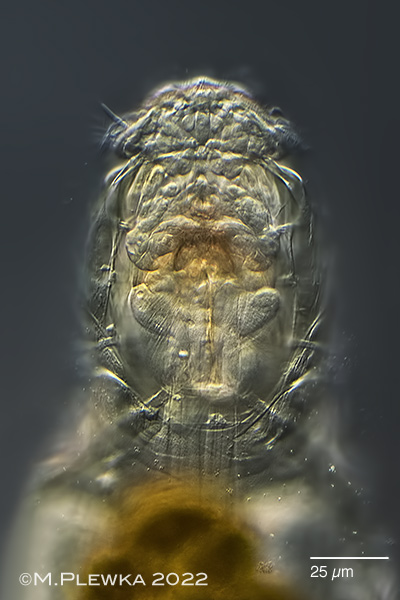 |
| Notommata copeus; characteristic for this species is the horsehead-like ventral ciliated field (buccal field, ventral view) (4). Right image: anterior part; dorsoventral view; optical median section (3) |
| |
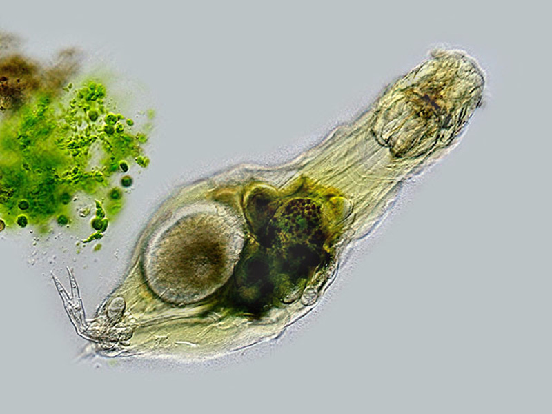 |
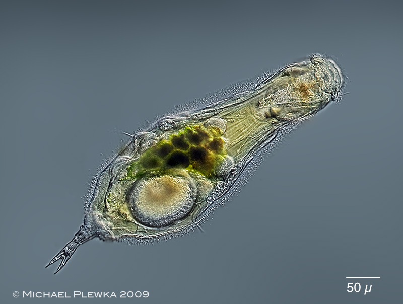
| Notommata copeus; swimming. The diet consists of filamentous algae and desmids. The green material is just digested and excreted Closterium algae. |
| |
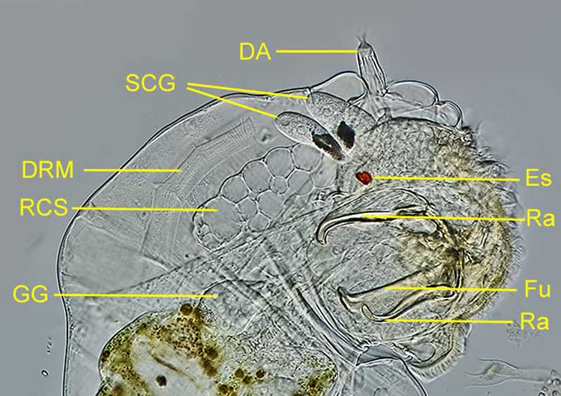 |
| Notommata copeus: lateral view of a compressed specimen. DA: dorsal antenna; DRM: dorsal retractor muscle for the head; ES: eyespot; Fu: fulcrum of the mastax Mx; Ra: Rami; RCS: retrocerebral sac. SCG: subcerebral glands with dark inclusions. |
| |
 |
 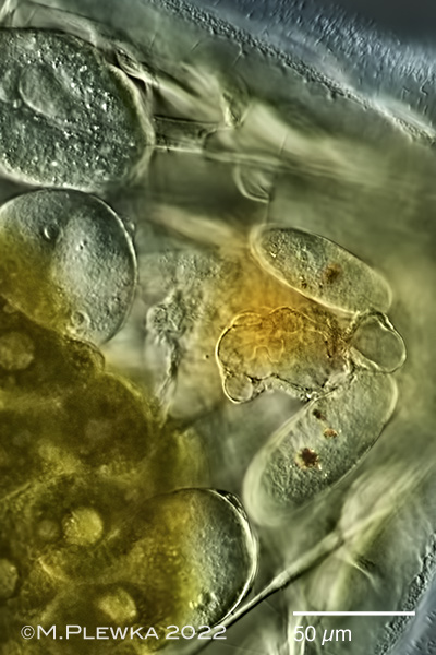 |
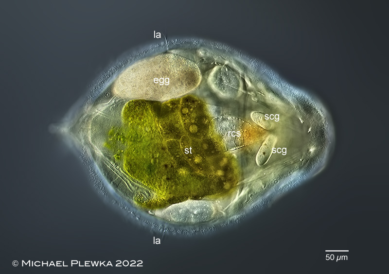
Notommata copeus: two images of the same specimen with comparison of the retrocerebral sac (RCS). Upper image: specimen with "filled" retrocerebral sac; head retracted. Lower left: crop of the above image. Lower right image: obviously the content of the RCS can be pressed out within seconds, as the EXIF-data of the photographs show. egg: (?amictic?) egg; la: lateral antenna; rcs: retrocerebral sac; scg: subcerebral gland; st: stomach. (3) |
| |
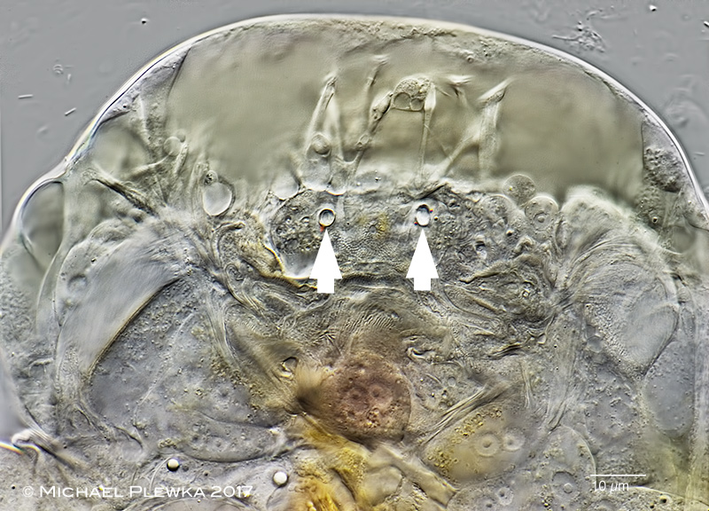 |
| Notommata copeus: dorsoventral view of anterior part of a compressed specimen with retracted corona. The arrowheads mark two structures that may be the openings of the ducts of the RCO. Similar structures have also been found in other Notommata-species, like for example Notommata allantois. |
| |
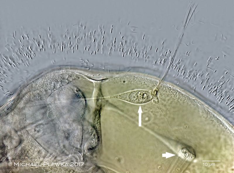 |
| Notommata copeus: detail of part of the posterior trunk; focus plane on the sensory cell of the lateral antenna (arrow) with sensory cilia and one of the retractor muscles (arrowhead). Also visible is the dense population of bacteria in the mucus layer surrounding the body. Maybe the mucus layer is a protection against chemical properties of the environment or a chemical "camouflage" against parasites. Also possible is a chemical signal for potential mating partners, but neither it is clear what function this mucus layer has, nor why the elongated bacteria are oriented perpendicular to the surface of the integument.(2). |
| |
  |
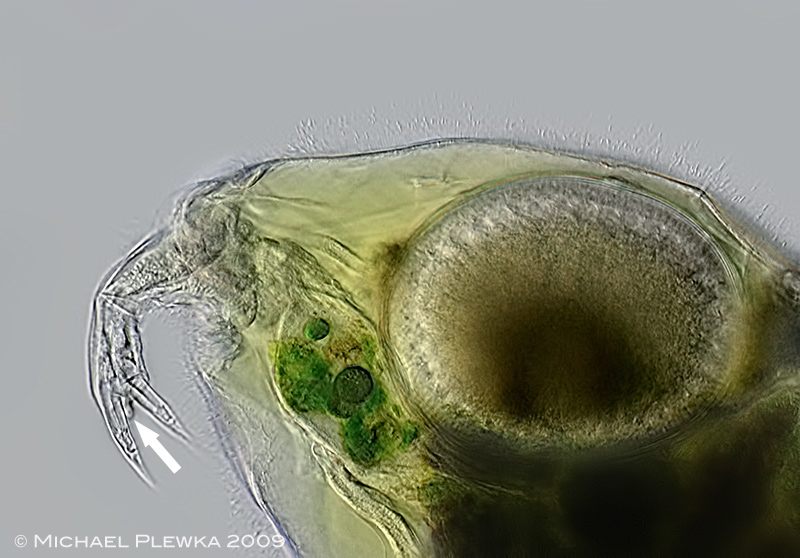
| Notommata copeus; left: detail of the (tip of the) lateral antenna, surrounded by bacteria in the mucus; right: appendage above the foot ("tail") which is typical for many Notommata species.. |
| |
 |
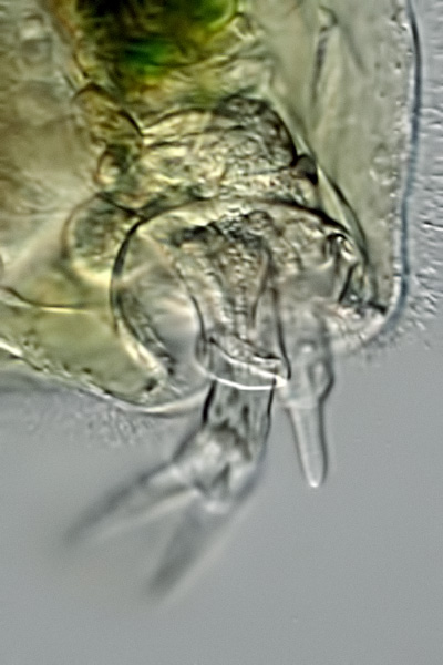
| Notommata copeus: the arrow points to a peg-like appendage between the toes.(2) |
| |
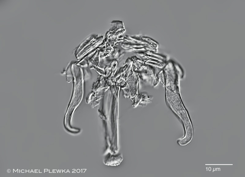 |
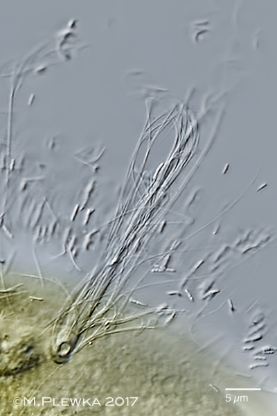
| Notommata copeus: virgate trophi.(2) |
| |
| |
| |
| |
| |
|
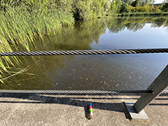
Location (3); : Henrichsteich, Hattingen, NRW, Germany, pond |
 |
| |
| Habitat (3); : plankton-sample with alga and detritus (click image to enlarge >>>) |
| |
| Date (3): 24.07.2022 |
| |
|
|
|
|
|
| |
| |
| |
| |
|
|
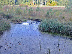 |
| Location(1); (2): Simmelried near Konstanz, BW, Germany, pond |
| |
| Habitat (1); (2); detritus (1); (2) |
| |
| Date : 22.7.2009 (1); 13.10.2017 (2) |
| |
|
|
|
|
|
| |
| |
| |
|
|
|