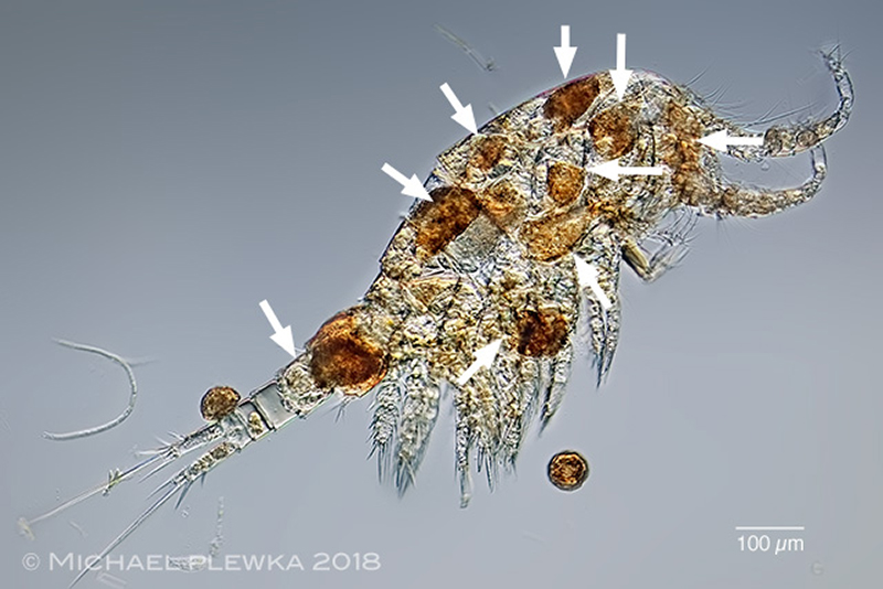
| Proales fallaciosa, may feed as a scavenger or is necrophagous, as seen here: the arrowheads point to 9 specimens feeding inside of the lorica of a copepod. (4). |
| |
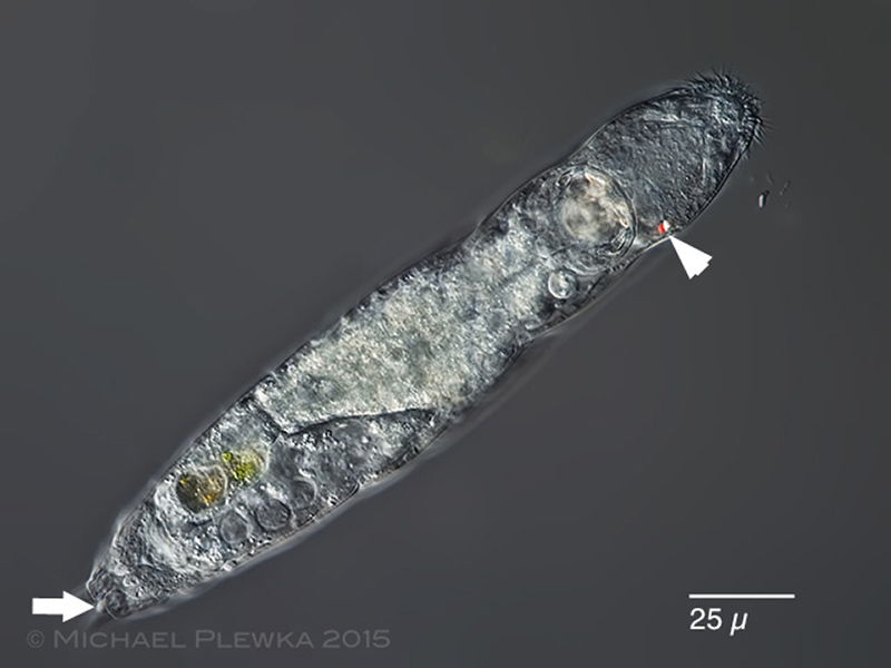 |
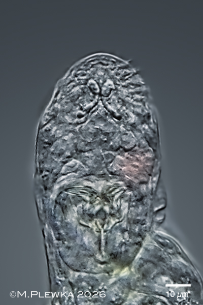
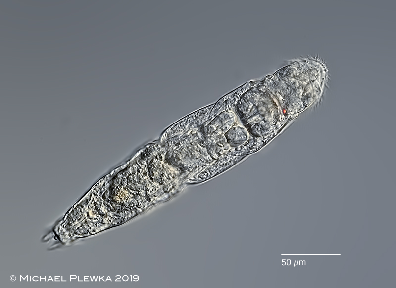
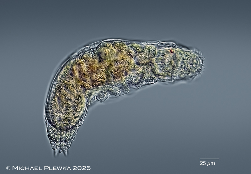
| Proales fallaciosa, dorsal view, optical transect. This species characterized by the single asymmetric orientated cerebral eyespot located on the right side (arrowhead) and a dorsal papilla/appendage between the toes (arrow; this appendage is in contrast to Proales decipiens) |
| |
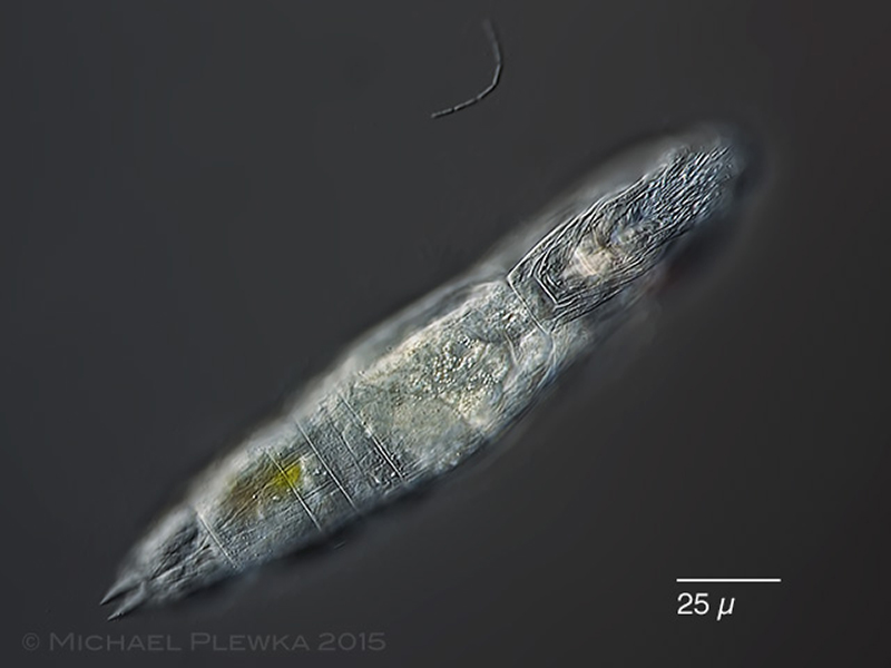 |
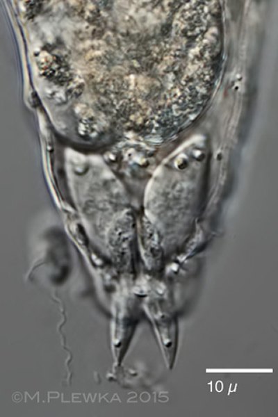
| Proales fallaciosa, ventral view of a specimen from (3). Focus plane is on the ventral ciliary field and the integument around the mouth (lower lip). |
| |
|
|
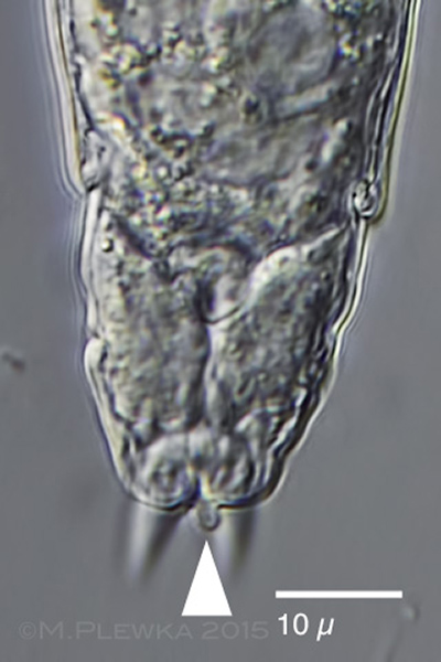
| Proales fallaciosa, two aspects of the foot of two specimens from (3). Left image: in contrast to Proales decipiens this species has a small dorsal appendix / papilla (arrowhead) between the toes. This may also lead to confusion with Notommata tripus, especially if the specimen is not fully expanded. Right: focus plane on toes and foot glands. |
| |
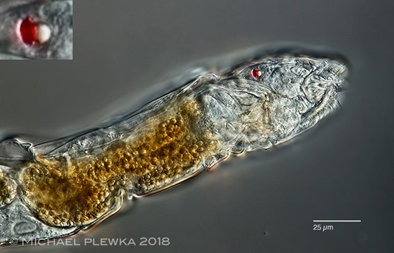 |
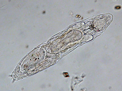
| Proales fallaciosa, lateral view of a specimen from (4). The insert shows the single eyespot which is displaced to the right part of the head. |
| |
| |
|
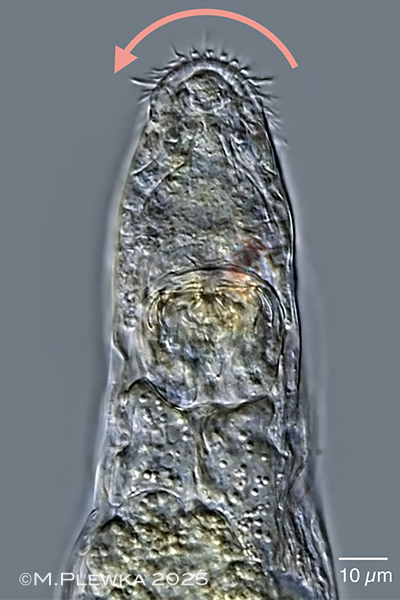
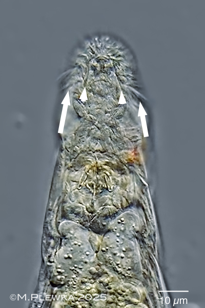
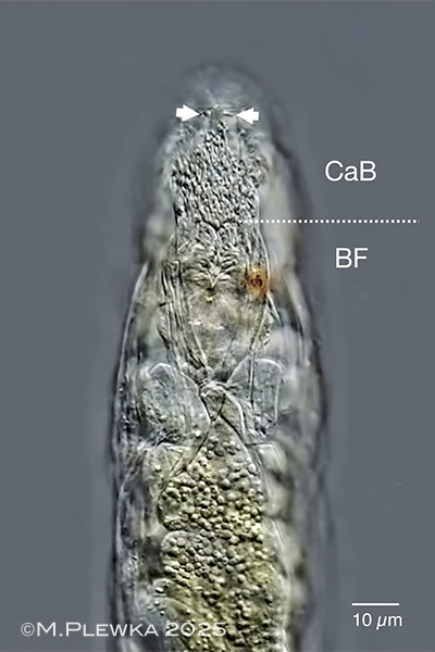
Proales fallaciosa, 4 optical transects in dorsoventral view of the anterior part. All 4 images are photographed from the dorsal side and are still images of a YT-video that shows the movement of the cilia of the corona.
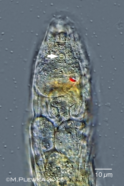
Upper left: focal plane on the dorsal antenna (arrowhead and the red eyespot. Also in focus are some tiny spots which are on the (undersurface of the ) coverslide. This shows that the dorsal side of the rotifer is orientated to the lens and not the ventral ciliated field and therefore the red eyespot is displaced to the right side and not to the left.
Upper right: the pink arrow indicates the direction of the metachronal movement of the locomotory cilia of this specimen under the given conditions: because this specimen is observed from the dorsal side, the metachronal movement of these cilia is anti-clockwise in this image. But when this specimen is observed from the front of the longitudinal axis (which is the reference orientation) the metachronal movement of these cilia would be clockwise. The effective stroke of these cilia, however, is directed backwards to the ventral side, so the metachronal movement is dexioplectic like in bdelloids or Gnesiotrocha.
Lower left: the row of motilie cilia (arrows) is separated by a membrane (arrowheads) from the cilia of the buccal field/ buccal tube.
Lower right: optical transect of the ventral side (the locomotory cilia are not in focus in this image). There are 3 different movements of the cilia which are visible here: the dotted line marks the border between the cilia of the circumapical band (CaB) and the cilia of the buccal field (BF). The metachronal movement of the cilia of the circumapical band (CaB) is symplectic, whereas the metachronal movement of the cilia of the buccal field (BF) is diaplectic and symmetric. The arrowheads mark two (??sensory??) cilia/ cirri which move transversely (the same can be observed in the rostral cilia of some Adineta_species). (7) |
| |
|
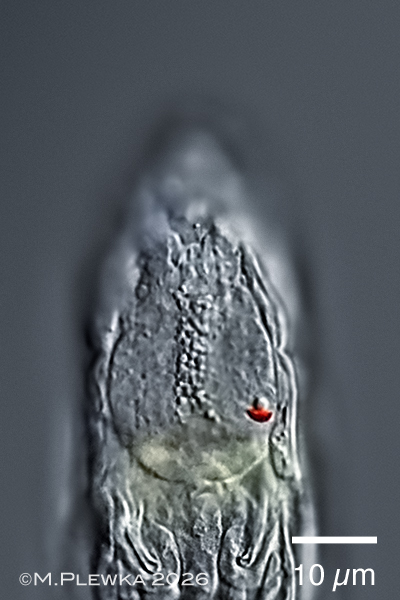
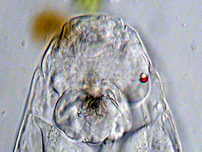
| Proales fallaciosa, anterior part, dorsal view. Left: focal plane on the displaced eyespot with lens and a granular structure that may be the duct of the retrocerebral sac. Right: optical median section, focal plane on the trophi and a structure that give the impression of eyespots, but may be actually the ducts of the subcerebral glands. (9) |
| |
| |
| |
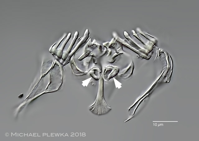 |
| Proales fallaciosa, virgate trophi; basal apophyses on rami; rami without alulae, which is in contrast to Proales sordida (4) |
|
| |
 |
| Proales fallaciosa, specimen from (5). |
| |
  |
| Proales fallaciosa, dorsoventral view. 2 images of specimen from (1) |
| |
| |
 |
| Proales fallaciosa; ventral! view of specimen from (8) |
| |
| |
|
|
| |
| |
|
|
| |
| |
|
|
| |
| |
|
|
| |
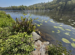
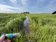
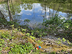
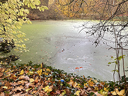
| Location: Gevelsberg Grünes Klassenzimmer, pond (2); Nature reserve NSG Schwalm, pond (3); Recker Moor, trench (4); Nature reserve NSG Heiliges Meer; Germany; Großes Heiliges Meer (5) |
| Habitat: periphyton (Utricularia) (3); detritus (4); epibiontic on the submerged leafs of Ranunculus fluitans (5). |
| Date: 06.11.2015 (3); coll: 13.06.2018, img.:23.06.2018 (4); 30.04.2019 (5) |
|
|