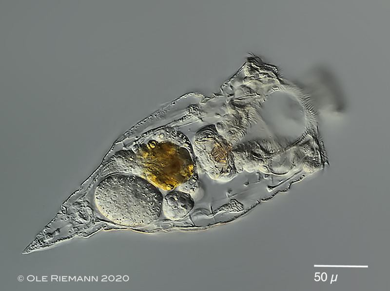
| Rhinoglena frontalis: female specimen, swimming, dorsoventral view. Specimen carrying a resting egg. (2) |
| |
 |
| Rhinoglena frontalis: another specimen, carrying an amictic egg. (2) |
| |
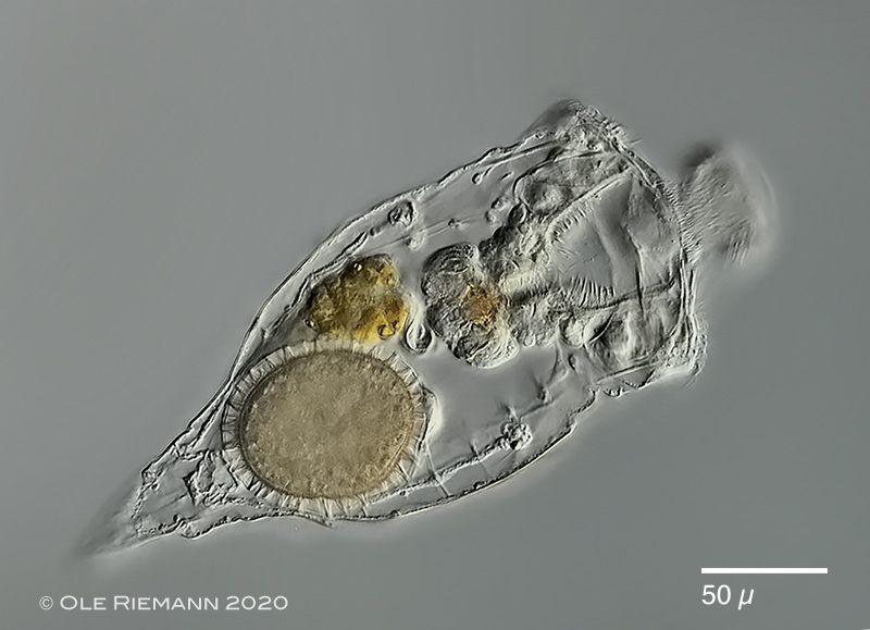 |
| Rhinoglena frontalis: another specimen with resting egg, which stays in the mother until she dies (Similar to some Lecane species). So in this species each female specimen can only produce a single resting egg during lifetime. (2) |
| |
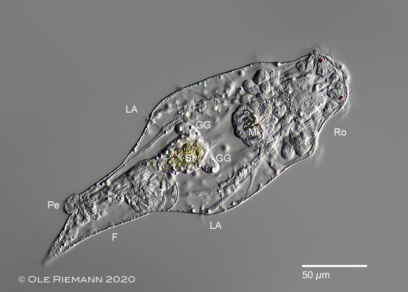 |
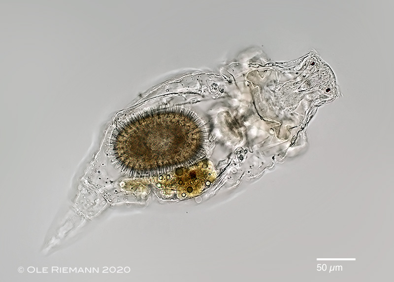
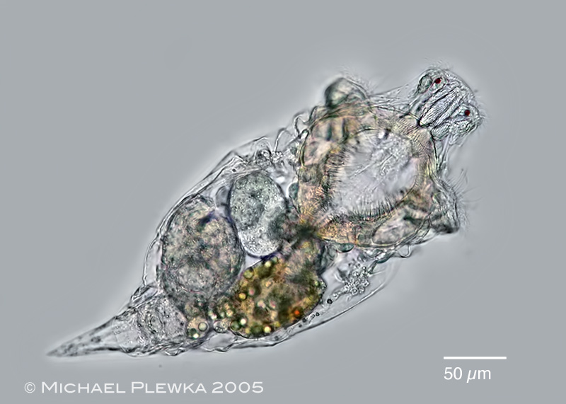
| Rhinoglena frontalis: male specimen. In contrast to the diploid females the males are haploid. While in most monogonont rotifers the males are so-called dwarf males, the males of Rhinoglena frontalis are only slightly smaller (see scale bar) than the females. In contrast to the males of other monogonont rotifers these males have a gut, comprising of: mastax (Mx) with trophi, stomach (St), gastric glands (GG). The green color of the content of the stomach ( i.e. green algae) indicates that the gut is fully functional. (2) |
| |
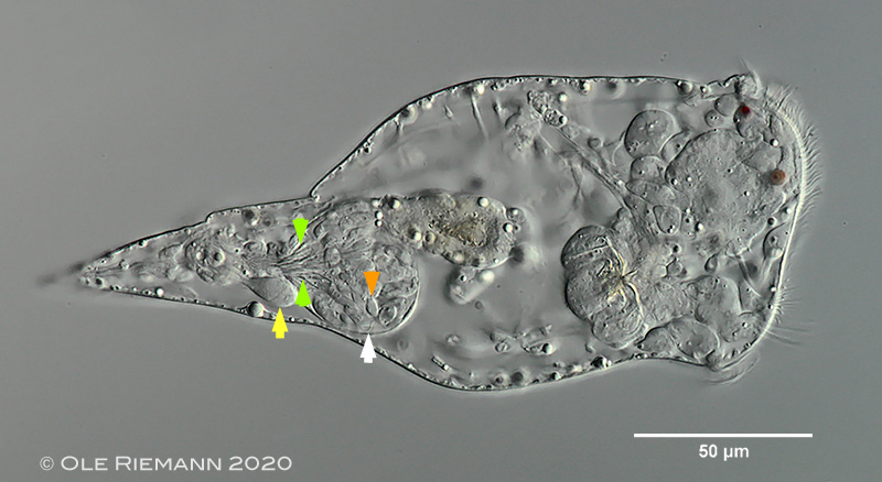 |
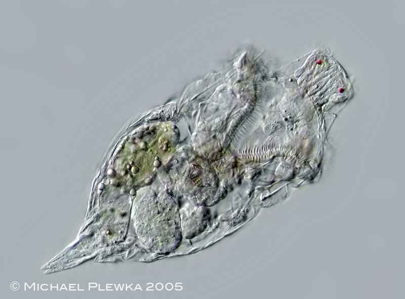
| Rhinoglena frontalis: another male specimen, slightly more compresssed than the specimen above. The orange arrowhead points to one of the elliptic sperm cells in the testis. The flagellum of the sperm cell is also visible (white arrowhead). The yellow arrowhead points to the accessory gland. The green arrowheads mark a bundle of needle-like structures, the so-called testicular rods.(2) |
| |
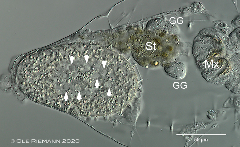 |
| Rhinoglena frontalis: slightly compressed resting egg of a female. The arrowheads point to some of the nuclei of the embryo. The presence of several nuclei indicates that the development of the embryo / cleavage has started already. Resting eggs can therefore be understood as diapausing embryos, which then will continue to develop after being activated by external stimuli. (2) |
| |
 |
 |
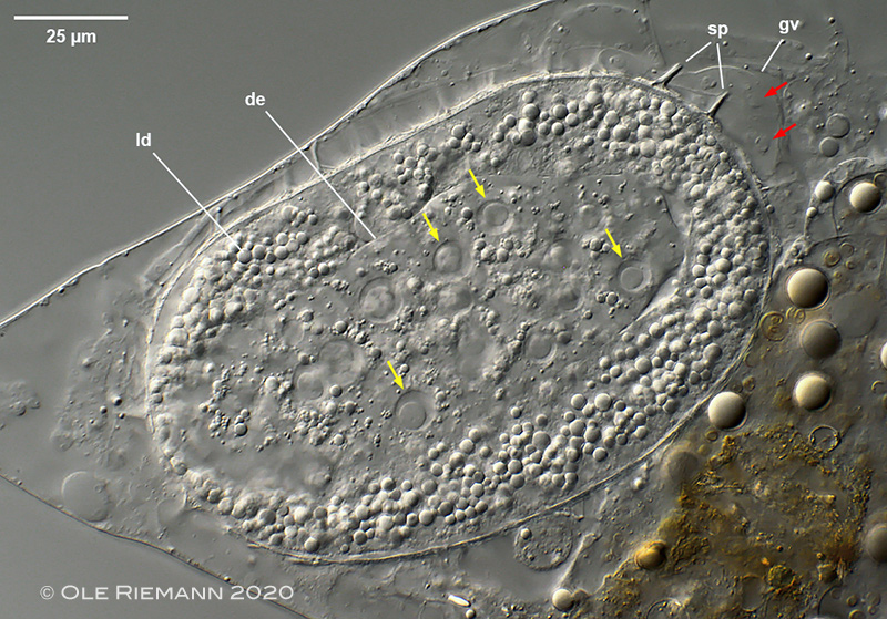
| Rhinoglena frontalis: images of 2 stages of the development of the resting egg: |
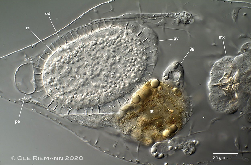
| upper image: 1. beginning of development. The red arrows point to the (degenerated) nuclei of the germovitellarium (gv); the yellow arrows point to some of the nuclei of the developing embryo (de). gv: germovitellarium; ld: lipid droplets. Two spines (sp) of the shell of the resting egg are already visible. (2) |
| lower image: 2. resting egg (re) with shell. The resting egg and the germovitellarium (gv) are enclosed by the oviduct (od). gg: gastric glands; pb: bladder; st: stomach (2) |
| |
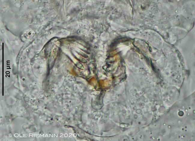 |
| Rhinoglena frontalis: malleate trophi of a female specimen. The trophi of the males are similar, but smaller. (2) |
| |
 |
| Rhinoglena frontalis: swimming specimen from (1). Ecology: R. frontalis is a cold-stenotherm plankton species. The population No. (1) was observed for one spring season in the pond of our school garden, which had been established 3 years before and still was nutritient-poor with few algae. In contrast to specimens of other rotifer species animals of R. frontalis were never seen again in the same pond as long as the pond existed (until 2015). Also the R. frontalis- specimens of location (2) were observed in a newly established pond with nutrient-poor water. (1) |
| |
 |
| Rhinoglena frontalis: swimming, ventral view, focus plane on the funnel-like mouth-opening (1) |
| |
| All images labelled (2) courtesy of Dr. Ole Riemann; University of Würzburg/ Germany. |
| |
| |
|
|
| |
| |
| |
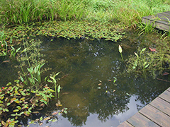
| Location: (2); Gerbrunn/ Würzburg, Germany; frog pond |
| Habitat (2): Plankton (2) |
| Date: 05.03.2020 (2) |
|
|